
Infected preauricular pits often present with localized pain and discharge, making it difficult for clinicians to determine the depth of the abscess without cross-sectional imaging. While these small indentations near the ear are frequently viewed as minor cosmetic issues, they represent the external opening of a potentially complex vestigial tract that can extend deep into the subcutaneous tissue. For many patients, the condition remains dormant for years, only to manifest as a painful, swollen mass during periods of physiological stress or bacterial exposure. In 2026, medical professionals increasingly recognize that relying solely on visual inspection often leads to incomplete treatment and high recurrence rates. The challenge lies in the unpredictable nature of the sinus path, which can branch or terminate near vital structures like the facial nerve. To address these complexities, clinicians have turned to high-resolution imaging to transition from a “wait-and-see” approach to a strategy of proactive, data-driven intervention that ensures better patient outcomes.
Using MRI for Diagnostic Precision
Identifying Structural Depth and Infection Markers
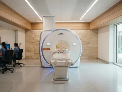
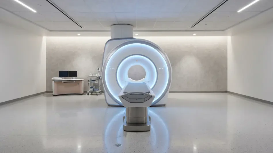
Magnetic resonance imaging has revolutionized the diagnostic process for preauricular anomalies by offering unparalleled soft-tissue contrast that highlights the exact extent of inflammatory processes. By utilizing specific protocols such as T2-weighted images with fat saturation, radiologists can isolate the fluid-filled tract from the surrounding adipose tissue with high specificity. This is particularly crucial during an active infection, where the presence of edema can obscure the physical boundaries of the sinus during a manual examination. These sequences allow the medical team to see the true length and orientation of the sinus, providing a three-dimensional understanding of how the infection has spread. Instead of guessing the depth of the tract based on the size of the cutaneous pit, surgeons can now rely on objective measurements to predict the complexity of the upcoming procedure.
Beyond standard anatomical imaging, the application of Diffusion-Weighted Imaging (DWI) provides a functional look at the nature of the lesion by identifying areas of restricted diffusivity. When purulent material or pus accumulates within a preauricular abscess, the movement of water molecules is significantly hindered, creating a distinct radiological signature that is easily identifiable. This allows clinicians to differentiate between simple cellulitis, which requires only antibiotic therapy, and a localized abscess that may necessitate immediate surgical drainage. In the clinical environment of 2026, this level of detail is essential for preventing the overuse of broad-spectrum antibiotics while ensuring that surgical intervention is timed perfectly to when it will be most effective. The objective evidence provided by DWI removes the ambiguity often associated with deep-seated infections that do not yet show visible fluctuance on the skin surface.
Mapping Proximity to Vital Anatomical Landmarks
The anatomical region surrounding the external ear is a dense crossroads of critical biological structures, including the parotid gland and the superficial temporal artery. High-resolution MRI sequences allow radiologists to map the sinus tract’s trajectory relative to these landmarks, ensuring that the surgeon knows exactly what to expect before making the first incision. In many complex cases, the tract can dive deep toward the parotid fascia or run parallel to the vascular supply of the scalp. By having a clear visual of these relationships, the surgical team can plan their approach to avoid accidental ligation of vessels or trauma to glandular tissue. This mapping is not merely a convenience; it is a safety protocol that minimizes post-operative complications like hematomas or salivary fistulas, which were more common before the widespread adoption of high-resolution cross-sectional imaging.
One of the most delicate considerations in preauricular surgery is the preservation of the facial nerve, specifically its mastoid segment and the branches that govern facial expression. Although the preauricular sinus is typically more superficial than the main trunk of the facial nerve, anatomical variations can place the tract in precarious proximity to nerve branches. Using axial Constructive Interference in Steady State (CISS) sequences, imaging specialists can visualize the relationship between the inflammatory tract and the neural pathways with millimetric precision. This “roadmap” is particularly valuable during revision surgeries where previous infections or surgeries have created scar tissue, distorting the usual anatomical planes. By identifying the safe margins around the facial nerve, the medical team can perform a more thorough excision of the sinus tract, significantly reducing the risk of nerve injury while maximizing the chances of a complete cure.
Understanding Development and Differential Diagnosis
Exploring Embryological Origins and Demographics
The formation of a preauricular sinus is rooted in the intricate dance of embryogenesis during the sixth week of gestation. As the external ear begins to take shape, six mesenchymal proliferations known as the “hillocks of His” arise from the first and second branchial arches to form the auricle. A sinus typically develops when there is an incomplete fusion or a failure of entrapment of the ectoderm during this rapid growth phase. This embryological perspective is vital because it explains why the tract is lined with squamous epithelium and is prone to the same issues as the skin, such as the accumulation of keratin debris. Understanding that this is a developmental failure rather than a simple infection helps clinicians explain the condition to patients as a structural anomaly that requires a structural solution rather than just temporary medication.
Demographic data show that while the preauricular sinus is found globally, its prevalence varies significantly based on ethnic and geographic factors. In Caucasian populations, the incidence remains relatively low at approximately one percent, whereas in some African and Asian regions, it can be as high as ten percent. In the year 2026, genetic research continues to explore why these tracts are often unilateral and predominantly found on the right side, though bilateral cases are more likely to be hereditary. Recognizing these patterns allows healthcare providers to identify patients who may have a familial predisposition to the condition. Furthermore, understanding the demographic context ensures that clinicians are more vigilant in populations where the condition is common, potentially identifying the pits during routine pediatric screenings before they have the chance to become symptomatic or infected.
Distinguishing Sinuses from Similar Pathologies
The diagnostic utility of MRI extends into the realm of differential diagnosis, where it helps distinguish a preauricular sinus from other congenital or acquired lesions. For instance, first branchial cleft anomalies often present in a similar location but have a much different relationship with the external auditory canal and the facial nerve. While a preauricular sinus is usually located superior and lateral to the parotid gland, branchial cleft cysts can be intimately associated with the glandular tissue itself. MRI provides the spatial resolution necessary to see these subtle differences in origin and termination, preventing diagnostic errors that could lead to inappropriate surgical techniques. Clear imaging ensures that the patient is not subjected to an overly aggressive surgery for a simple sinus or an inadequate surgery for a more complex branchial anomaly.
Clinicians must also differentiate the sinus from common skin-related pathologies like epidermal inclusion cysts or trichilemmal cysts. On the surface, an infected inclusion cyst might look identical to an infected preauricular pit, especially if the pit is small or hidden by inflammation. However, MRI sequences can reveal the presence of the sinus tract extending toward the auricular cartilage, a feature that is absent in simple cysts. Additionally, the lack of internal calcifications—often seen in trichilemmal cysts—further narrows the diagnosis. By confirming the diagnosis through imaging, the medical team can avoid the frustration of “recurrent cysts” that are actually symptoms of an untreated sinus tract. This precision in 2026 ensures that the definitive treatment plan is established early in the patient’s journey, reducing the need for multiple, unsuccessful minor procedures.
Refining Surgical and Medical Management
Transitioning from Acute Infection to Surgery
Managing a symptomatic preauricular sinus requires a careful balance between addressing immediate inflammation and planning for long-term resolution. When a patient presents with an acute infection, the priority is to control the bacterial load and alleviate pain, typically through a course of antibiotics such as amoxicillin-clavulanate. MRI is often utilized during this phase to determine if an abscess has formed, which would necessitate incision and drainage. However, the definitive cure for a preauricular sinus is not found in medicine but in surgery. The goal is to move the patient into the “quiescent phase”—a period where the infection has subsided, and the tissue is no longer friable. During this calm window, the sinus tract is most clearly defined on imaging, making it the ideal time for a successful surgical excision that targets the entire epithelial lining.
The transition from medical management to surgery is where MRI provides its greatest value as a planning tool. Once the acute swelling has decreased, high-resolution scans can be performed to capture the resting state of the sinus. This allows the surgeon to visualize the tract without the distortion of active edema, ensuring that every branch of the “fistula auris” is accounted for. This pre-operative visualization is essential because any remaining epithelial cells can lead to a recurrence of the infection, often with more severity than the original presentation. In 2026, the standard of care increasingly dictates that surgery should only be performed once the anatomy is fully understood through imaging, moving away from the older practice of “blind” exploration that frequently resulted in incomplete removals and multiple return visits to the operating room.
Selecting the Optimal Surgical Technique
Surgical success in treating a preauricular sinus depends heavily on choosing the right approach for the patient’s specific anatomy as revealed by MRI. The two most common techniques are the simple sinectomy and the supra-auricular approach. A simple sinectomy involves an elliptical incision around the pit and the dissection of the tract, but it carries a higher risk of recurrence if the tract is deep or branched. In contrast, the supra-auricular approach involves a wider dissection that identifies the temporalis fascia as a deep boundary, allowing for a more comprehensive removal of the affected tissue. By reviewing the MRI data beforehand, a surgeon can determine if the tract’s complexity warrants the more extensive supra-auricular method, thereby tailoring the invasiveness of the procedure to the actual needs of the individual patient.
The data provided by MRI also help surgeons minimize the use of post-operative drains, which can sometimes serve as a nidus for new infections. When a surgeon has high confidence in the complete excision of the tract based on a clear radiological “roadmap,” they can often achieve a cleaner closure with less risk of dead space where fluid might accumulate. This precision leads to faster recovery times and better cosmetic results, as the incisions can be smaller and more strategically placed. Furthermore, by avoiding vital structures identified on the MRI, the surgeon can perform the excision with greater speed and less blood loss. This evolution in surgical strategy highlights how advanced imaging has transformed a once-routine minor surgery into a sophisticated, targeted intervention that prioritizes long-term patient health and the prevention of chronic, recurring symptoms.
Determining the Need for Systemic Screening
The presence of a preauricular sinus is not always an isolated finding; it can sometimes be a marker for broader congenital issues, particularly those affecting the renal system. In 2026, the decision to pursue systemic screening, such as a renal ultrasound, is guided by the “Wang guidelines,” which look for specific risk factors in the patient’s history and physical presentation. If the MRI reveals a particularly complex or bilateral sinus, or if there is a family history of hearing loss or maternal gestational diabetes, the suspicion for a syndromic condition like Branchio-Oto-Renal (BOR) syndrome increases. By using the imaging results as part of a holistic diagnostic picture, clinicians can ensure that they are not treating the ear in a vacuum while overlooking potentially serious internal malformations.
Building on the diagnostic insights gained from the head and neck imaging, clinicians can more accurately counsel families about the implications of the congenital pit. If the MRI shows a simple, unilateral tract and there are no other dysmorphic features, the medical team can provide reassurance that the risk of associated anomalies is low, avoiding unnecessary and costly systemic tests. Conversely, when imaging suggests a more involved developmental failure, the team can proactively coordinate with specialists in nephrology or audiology. This integrated approach ensures that the preauricular sinus is treated with the appropriate level of clinical concern. By leveraging technology to separate isolated cases from systemic syndromes, modern medicine provides a more efficient and personalized healthcare experience that addresses the root cause of the patient’s condition while safeguarding their overall well-being.
Evolution of Management Strategies and Outcomes
The integration of high-resolution MRI into the treatment of preauricular sinuses was a significant advancement that improved the precision of surgical interventions. By providing a detailed map of the internal tract and its relationship to the facial nerve and parotid gland, imaging allowed surgeons to move beyond superficial visual cues. This shift resulted in a measurable decrease in recurrence rates, as surgeons were better equipped to remove every remnant of the epithelial lining. The use of functional sequences like DWI further refined the timing of these procedures, ensuring that operations were performed during the most favorable clinical windows. Ultimately, the transition to an imaging-first approach simplified the management of a common but traditionally stubborn congenital anomaly.
Future clinical care should focus on the standardization of imaging protocols for all symptomatic preauricular anomalies to ensure consistent outcomes across different healthcare settings. Practitioners are encouraged to adopt the multiplanar MRI sequences discussed to provide a comprehensive roadmap for every patient facing recurrent infections. Additionally, the use of targeted surgical approaches guided by these scans should become the baseline for managing complex or revision cases. By continuing to bridge the gap between radiological detail and surgical execution, the medical community can virtually eliminate the cycle of chronic recurrence that once plagued patients with this condition. Prioritizing this level of diagnostic depth will lead to more efficient recovery times and a higher standard of permanent resolution for congenital ear defects.