
While thyroid incidentalomas found on PET/CT scans carry a historical malignancy risk of up to 54%, metabolic data often fails to provide a diagnostic advantage over standard ultrasound. This diagnostic dilemma has long complicated the clinical management of patients undergoing whole-body imaging for nonthyroidal malignancies. When a PET scan reveals a “hot” focal area in the thyroid gland, the immediate reaction is often one of high clinical concern, leading many practitioners to prioritize metabolic activity over established anatomical risk assessments. However, a comprehensive retrospective study conducted at Samsung Medical Center has challenged this reliance by evaluating whether adding metabolic metrics truly improves diagnostic accuracy. The study aimed to determine if fluorine-18 fluorodeoxyglucose (FDG) uptake could refine the predictive power of systems like the American College of Radiology Thyroid Imaging Reporting and Data System (ACR-TIRADS). By analyzing a high-risk cohort from 2026 perspectives, the research clarifies the hierarchy of imaging modalities in thyroid care.
Study Population and Methodological Rigor
Assessing Clinical Data and Patient Demographics
The study cohort was derived from a major tertiary referral center, a factor that naturally resulted in a high baseline prevalence of thyroid malignancy among the participants. Out of the 464 nodules analyzed, approximately 47.6% were eventually confirmed as malignant through pathological testing, while 52.4% were determined to be benign. This nearly balanced distribution provided a statistically robust dataset for evaluating the nuances of imaging performance. It is important to note that the demographic was predominantly composed of patients already battling other forms of cancer, with nearly 97% of the subjects undergoing PET/CT for staging or surveillance of nonthyroidal primary tumors. This specific patient profile means the results are particularly relevant for oncology specialists managing complex cases where multiple suspicious findings may emerge simultaneously during routine diagnostic workups.
The researchers focused on patients who had undergone a comprehensive battery of tests between 2026 and earlier periods, ensuring each case had definitive results from neck ultrasonography and biopsy. This rigorous selection process was designed to eliminate ambiguity, excluding patients who had non-diagnostic cytology or a history of previous thyroid surgery that could distort imaging results. By focusing on a population where the likelihood of finding a secondary malignancy is inherently higher, the study provided a stress test for current diagnostic frameworks. The high prevalence of malignancy in this specific group underscores the necessity for diagnostic tools that are not only sensitive but also highly specific to avoid unnecessary invasive interventions in patients who are already undergoing intensive treatments for other serious health conditions.
Standardizing Imaging and Quantitative Protocols
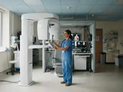
To maintain the highest level of data integrity, the researchers implemented strict standardization for all imaging protocols used during the study period. Patients were required to fast for at least six hours prior to their PET/CT scans to ensure that blood glucose levels remained below 200 mg/dL. This precaution is vital because high blood sugar can cause competition between natural glucose and the radiotracer for cellular uptake, which would artificially lower the observed metabolic activity in a nodule. The PET/CT scans themselves were performed approximately 60 minutes after the administration of 5.0 MBq/kg of 18F-FDG. Such precision ensured that the captured metabolic data was a true reflection of the cellular activity within the thyroid nodules, rather than a byproduct of physiological variations or technical inconsistencies during the scanning process.
The quantitative analysis of these scans was handled by two experienced nuclear medicine specialists who were blinded to the final pathological outcomes of the patients. They focused on calculating the maximum standardized uptake value (SUVmax) using three-dimensional volumes of interest, providing a consistent numerical baseline for comparison. Simultaneously, ultrasound evaluations were conducted within a median of 21 days from the PET/CT, ensuring that the anatomical and metabolic snapshots were temporally aligned. This close alignment is critical for accurate risk stratification, as thyroid nodules can change in size or appearance over longer durations. By ensuring that the two imaging modalities reflected the same stage of disease progression, the study was able to provide a direct and fair comparison of how each technology contributes to the final diagnostic decision.
Integration of Metabolic and Anatomical Data
Evaluating Ultrasound as the Primary Modality
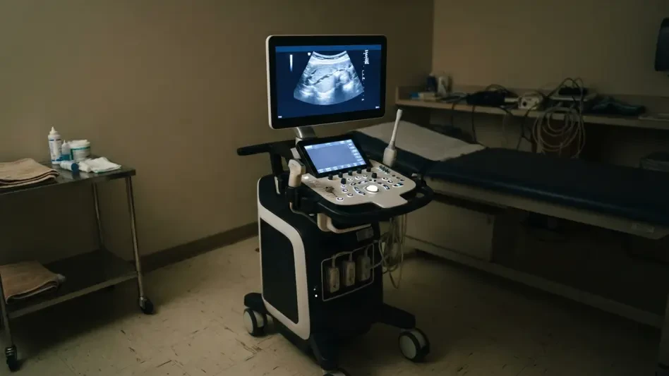
Ultrasonography continues to serve as the frontline tool for evaluating thyroid health, utilizing sophisticated scoring systems to categorize the risk of malignancy. In this study, systems like ACR-TIRADS and the Korean K-TIRADS were used to analyze nodules based on features such as echogenicity, shape, margins, and the presence of microcalcifications. The results reaffirmed the incredible sensitivity of these anatomical frameworks, with approximately 90% of all confirmed malignant nodules already being classified in the moderately or highly suspicious tiers by ultrasound alone. This baseline efficiency highlights the significant challenge for any adjunctive imaging modality: to be truly useful, it must provide a clear benefit that ultrasound does not already offer through its detailed visualization of nodule structure and composition.
When the researchers attempted to integrate focal FDG uptake as a binary variable into the ultrasound-based models, the results were statistically significant but clinically underwhelming. The Area Under the Receiver Operating Characteristic (AUROC) curve, which measures a test’s overall ability to distinguish between disease and health, showed only a marginal increase for both the ACR and Korean systems. More importantly, the Net Reclassification Improvement (NRI) was statistically insignificant, meaning the addition of metabolic data did not change the risk category of most nodules in a way that would alter recommended clinical management. These findings suggest that for the vast majority of patients, the structural information provided by a high-quality ultrasound is already sufficient for making an accurate diagnosis without the need for additional metabolic confirmation.
The Reclassification Paradox and Diagnostic Penalties
The study revealed a complex “reclassification paradox” when metabolic data was used to supplement traditional ultrasound findings. There was a notable improvement in the “non-event NRI,” which refers to the ability of the imaging model to correctly identify benign nodules. In cases where a nodule showed no metabolic activity on a PET scan, the model became more confident in labeling it as benign, which could theoretically spare some patients from undergoing an unnecessary fine-needle aspiration. This potential to reduce overdiagnosis is an attractive feature in modern medicine, where avoiding low-value invasive procedures is a high priority. However, this gain was entirely offset by a significant failure in the “event NRI,” which tracks the accuracy of identifying confirmed malignant cases.
This diagnostic failure represented a major “penalty” for clinicians relying too heavily on PET data. The integrated models inappropriately down-classified several truly malignant nodules simply because they did not exhibit high radiotracer uptake. In the context of oncology, a false negative is a far more dangerous outcome than a false positive, as it can lead to delayed treatment for a growing cancer. Using a lack of metabolic activity to “downgrade” the risk of a nodule that already looks suspicious on an ultrasound introduces an unacceptable level of risk. The data clearly indicated that “metabolic silence” is not a guarantee of safety. Consequently, the research emphasized that while PET might offer some reassurance for low-risk nodules, it should never be used to override the warning signs found during a standard sonographic examination.
Quantitative Analysis of SUVmax
Investigating Intensity Thresholds and Clinical Benefit
Moving beyond the simple presence of radiotracer uptake, the researchers investigated whether the intensity of that uptake, measured by the SUVmax, could provide more definitive diagnostic clarity. The data showed that malignant nodules indeed had a significantly higher mean SUVmax compared to benign ones, suggesting a correlation between high metabolic activity and the presence of cancer. However, the fundamental problem remained the absence of a reliable cutoff value. There was no specific SUVmax threshold that could effectively separate benign and malignant nodules with enough certainty to guide clinical decisions. Even at lower levels of metabolic activity, the risk of malignancy remained high enough that a biopsy would still be required if the ultrasound appearance was concerning.
Further complicating the issue was the result of the Decision Curve Analysis (DCA), which evaluates the net clinical benefit of a diagnostic test at different probability thresholds. The analysis showed that adding quantitative SUVmax data only provided a benefit when the probability of malignancy was estimated to be above 45%. In actual clinical practice, the decision to perform a biopsy is typically made at much lower thresholds, often when the risk is as low as 5% to 20%. Because the “added value” of the PET data only appears at high-risk levels where a biopsy would already be mandatory, it fails to provide any meaningful guidance in the “gray zone” where clinicians need the most help. Thus, quantitative metabolic data often serves as a redundant confirmation of risk rather than a tool that changes the course of patient care.
Pathological Diversity and Distribution
The study benefited from a remarkably diverse range of thyroid pathologies, which is a hallmark of the complex cases handled at major oncology centers. While the majority of the malignant cases were identified as papillary thyroid carcinomas, the researchers also documented several instances of much more aggressive diseases. These included thyroid lymphoma, anaplastic thyroid carcinoma, and metastatic lesions originating from distant primary cancers like the lungs or the colon. These high-grade malignancies typically exhibited very high metabolic activity, which accounted for the high average SUVmax seen in the malignant group. The presence of such diverse and dangerous pathologies highlights the importance of having a robust diagnostic strategy that can capture a wide spectrum of cellular behaviors.
In contrast, the “false negative” results on the PET scans were frequently associated with conventional papillary thyroid carcinomas. These tumors, while definitely malignant, can be metabolically indolent, meaning they do not consume glucose at a rate significantly higher than surrounding healthy tissue. This creates a dangerous situation where a life-threatening cancer might appear “quiet” on a metabolic scan while showing clear structural irregularities on an ultrasound. This discrepancy is a primary reason why metabolic imaging is an inconsistent tool for thyroid assessment. The biological diversity of thyroid cancer means that some of the most common forms of the disease can easily evade detection if clinicians rely solely on PET data, further solidifying the role of ultrasound as the more reliable primary diagnostic modality.
Clinical Implications and Expert Recommendations
Prioritizing Ultrasound Over Metabolic Silence
The central finding of this investigation is that clinicians must prioritize anatomical features over metabolic silence when managing thyroid nodules. The research provided unequivocal evidence that the absence of focal FDG uptake is not a “safety net” that can be used to avoid a biopsy. Malignancy rates remained alarmingly high—reaching up to 77.8%—for nodules that lacked metabolic avidity but were classified as highly suspicious under K-TIRADS criteria. These statistics serve as a stark warning to any medical professional who might be tempted to delay a biopsy based on a negative PET scan. The structural characteristics of a nodule, such as its shape, margins, and internal composition, remain far more predictive of its true nature than its glucose metabolism.
Building on these findings, the recommended clinical workflow in 2026 emphasizes that if a thyroid nodule meets the size and sonographic criteria for a biopsy according to standard TI-RADS guidelines, that procedure must be performed regardless of the PET scan results. The “glowing” or “silent” status of a nodule on a PET/CT should be viewed as secondary information that does not override the gold-standard anatomical assessment. This approach ensures that patients with metabolically indolent but structurally suspicious cancers receive the timely intervention they need. By maintaining a strict adherence to ultrasound-based risk stratification, clinicians can minimize the risk of missing aggressive cancers that do not conform to typical metabolic patterns, thereby improving overall patient safety and diagnostic accuracy.
Acknowledging Study Limitations and Context
The researchers were transparent about several limitations that must be considered when applying these results to broader clinical settings. One major factor was the potential for verification bias, as the study was conducted at a tertiary cancer center where patients were more likely to have malignancies than the general population. In a community health setting where patients are typically healthier, the malignancy rate of thyroid incidentalomas might be lower, which could theoretically improve the negative predictive value of metabolic imaging. Furthermore, the retrospective nature of the study meant that the imaging protocols were not originally designed for a head-to-head comparison, and the use of different PET scanner models without cross-calibration could have introduced minor variations in the SUVmax measurements.
The final synthesis of the research demonstrated that while PET/CT is an invaluable tool for systemic oncology, its role in localized thyroid nodule evaluation is largely supplementary. The medical community has recognized that the initial hype surrounding metabolic imaging for thyroid nodules must be balanced with a realistic understanding of its limitations. The study proved that structural ultrasound remains the most effective gatekeeper for deciding which nodules require invasive testing. Ultimately, the integration of metabolic data should only be considered an adjunct to, never a replacement for, high-quality sonographic analysis. By recognizing these boundaries, the medical field has moved toward a more evidence-based diagnostic pathway that prioritizes anatomical precision over metabolic activity, ensuring that patient management remains both safe and efficient in the face of complex incidental findings.