
The integration of the Adaptive Image Noise Optimizer allows for significant tube current reduction without losing the bone-to-soft-tissue clarity required for patient-specific implants. This technological advancement represents a critical pivot in the management of complex congenital conditions, where the precision of surgical outcomes must be balanced against the biological risks of cumulative radiation. In the specialized field of cleft and craniofacial surgery, practitioners at the Cleft Palate and Craniofacial Center at Helsinki University Hospital (HUSUKE) have spearheaded research that validates the transition from standard-dose protocols to ultra-low-dose (ULD) cone-beam computed tomography (CBCT). By evaluating the efficacy of these protocols in patients requiring intricate reconstructions, the medical community is finding that the historical reliance on high-dose imaging is no longer a prerequisite for surgical excellence. As pediatric and adolescent patients often face a long-term trajectory of corrective procedures, the ability to obtain high-fidelity three-dimensional data at a fraction of the traditional radiation cost is not merely a convenience but a fundamental improvement in patient safety standards. This shift is particularly relevant as the industry moves toward more personalized, data-driven surgical workflows that utilize virtual planning and 3D printing to achieve anatomical harmony in even the most challenging cases.
Comparing Standard and Low-Dose Protocols
Clinical Framework: Study Demographics and Imaging Targets
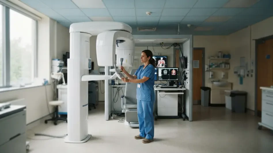
The study conducted at HUSUKE analyzed a cohort of 40 patients between 2026 and 2028, reflecting a diverse range of craniofacial needs including unilateral and bilateral cleft lip and palate, along with other rare congenital anomalies. These patients, ranging in age from early adolescence to young adulthood, underwent a rigorous imaging comparison between the established FACE protocol and the newer ULD JAW protocol. The FACE protocol, designated as setting F2 on the Planmeca Viso G7 scanner, served as the standard-dose benchmark, while the JAW protocol, setting J13, represented the ultra-low-dose alternative. The primary objective was to determine if the JAW protocol could provide a sufficient foundation for virtual surgical planning (VSP) without the typical degradation in image quality associated with lower tube currents. By focusing on patients who were already scheduled for orthognathic or reconstructive surgery, the researchers were able to test the ULD protocol in a high-stakes clinical environment where the margin for error in bone segmentation is virtually nonexistent. This demographic focus is essential because the adolescent jaw is a dynamic structure, and the precision of the resulting digital models directly influences the long-term success of the reconstruction.
Digital Planning: The Path from DICOM to Model
Navigating the transition from raw DICOM data to a finalized patient-specific implant (PSI) requires a sophisticated digital workflow that bridges the gap between radiological science and surgical engineering. In this study, the processing of CBCT data was handled using Planmeca Romexis software, which allowed for the initial visualization and preparation of the volumetric datasets. Following this, the data was exported for virtual planning in Geomagic Freeform, a platform known for its ability to handle complex anatomical structures with high fidelity. The bone segmentation process, which isolates the hard tissue from the surrounding soft tissue and electronic noise, was performed by planning specialists with over 15 years of experience. This high level of human expertise was coupled with the Adaptive Image Noise Optimizer (AINO) to ensure that the resulting Standard Tessellation Language (STL) models were accurate representations of the patient’s anatomy. The success of this workflow was measured by the intraoperative fit of cutting guides and implants, which were assessed by surgeons based on their anatomical seating and stability. If a guide did not rock and required no manual modification, the imaging protocol was deemed successful, confirming that ULD data can indeed support the manufacturing of precision-grade surgical tools.
Assessing Radiation and Surgical Outcomes
Exposure Reduction: Analyzing the Radiation Metrics
The quantitative results of the study underscore a massive leap forward in the application of the ALARA (As Low As Reasonably Achievable) principle within the operating room. The researchers measured the dose-area product (DAP) and calculated the effective dose for each patient, revealing that the ULD JAW protocol achieved a median effective dose of only 40.3 µSv. In contrast, the standard-dose FACE protocol resulted in a median dose of 89.0 µSv, representing a reduction of more than 50 percent. This reduction is transformative because it places the radiation exposure of a full 3D volumetric dataset in the same range as traditional 2D dental X-rays, which provide significantly less diagnostic information. For a patient born with a cleft condition who might require dozens of scans over a 20-year treatment window, this cumulative reduction drastically lowers the lifetime risk of radiation-induced complications. Furthermore, the ability to maintain diagnostic integrity at 40.3 µSv demonstrates that modern hardware and software can effectively compensate for lower photon counts, allowing clinicians to prioritize patient safety without compromising the anatomical detail necessary for complex bimaxillary reconstructions and Le Fort I osteotomies.
Surgical Precision: Evaluating Intraoperative Accuracy
While the reduction in radiation is a clear victory for patient safety, the ultimate test of the ULD protocol lies in the surgical outcomes and the physical fit of the implants produced from the data. The HUSUKE study found no statistically significant difference in the intraoperative performance of cutting guides and patient-specific implants between the ULD and standard-dose groups. Out of the 40 implants evaluated, 37 were deemed to have a perfect fit upon initial placement, requiring no adjustments to the bone or the implant itself. In the few cases where minor misfits occurred, they were linked to mechanical factors such as screw hole alignment or physical constraints within the surgical site, rather than deficiencies in the underlying CBCT image quality. This high success rate across both groups proves that the ULD JAW protocol provides a reliable foundation for even the most demanding orthognathic movements. Surgeons reported that the stability of the guides was consistent, and the resulting occlusion and facial symmetry met all preoperative goals. This evidence suggests that the medical community can safely move away from high-dose scans, as the “noise” in ultra-low-dose images does not translate into physical inaccuracies in the final 3D-printed surgical hardware.
Analyzing Clinical Decisions and Industry Trends
Selection Factors: The Role of Patient Demographics
A notable finding in the research was the specific set of factors that influenced which patients were assigned to the ultra-low-dose protocol during the study period. Through binary logistic regression analysis, the researchers determined that patient age was the most significant predictor for the use of the ULD JAW protocol. Clinicians were instinctively more inclined to utilize lower radiation settings for younger patients, reflecting a proactive clinical effort to minimize lifetime exposure in a demographic that is more sensitive to ionizing radiation. Interestingly, other variables such as the severity of the cleft, the presence of other craniofacial deformities, or the specific type of surgery (e.g., unilateral vs. bilateral) did not significantly impact the choice of protocol. This indicates that the ULD protocol is increasingly viewed not as a specialized tool for “easy” cases, but as a versatile standard that can be applied across the board. The fact that the protocol performed equally well regardless of the patient’s complexity suggests that the industry is ready to adopt ULD as a primary imaging modality, moving beyond its initial role as a niche alternative for pediatric patients.
Digital Twins: The Future of Virtual Planning
The broader trend identified in the study is the rapid shift toward the use of “Digital Twins” in maxillofacial surgery, where a perfect digital replica of the patient’s anatomy is used to simulate every step of the procedure before the first incision is made. As of 2026, the reliance on virtual surgical planning has made the quality of bone segmentation the most critical step in the reconstructive chain. The study highlights that modern reconstruction algorithms like AINO have reached a level of maturity where they can effectively “clean” low-dose images, making them indistinguishable from high-dose images for the purpose of 3D modeling. Because the fit of an implant is an integrated outcome—influenced by 3D printing tolerances, the expertise of the planning technician, and the tension of the patient’s soft tissues—the slight variations in raw image noise from ULD scans are essentially filtered out during the multi-step production process. This realization is pushing the industry to focus more on the software’s ability to interpret data rather than simply increasing the power of the hardware. As these digital workflows become more automated, the integration of low-dose imaging will likely become the baseline requirement for any facility performing high-precision craniofacial reconstructions.
Future Research and Clinical Standards
Research Context: Recognizing Study Constraints
Despite the overwhelmingly positive results, the study authors acknowledged several limitations that must be addressed to further solidify these findings within global clinical guidelines. The research was conducted at a single-center facility, HUSUKE, which is known for its high level of specialization and access to state-of-the-art equipment like the Planmeca Viso G7. This raises the question of whether similar results can be achieved at facilities with different hardware or less experienced planning teams. Additionally, the sample size of 40 patients, while sufficient for a pilot validation, needs to be expanded in multicenter trials to account for a wider variety of surgical techniques and ethnic anatomical variations. Another point of discussion involves the qualitative nature of the implant fit assessment. While the surgeons’ expert observations are the most practical metric for intraoperative success, the researchers refrained from taking postoperative CT scans to quantitatively measure the exactness of bone movement. This decision was made to strictly adhere to the ALARA principle, highlighting the ethical dilemma of modern radiological research: validating safety and accuracy without subjecting the subjects to the very radiation the study seeks to minimize.
Advancing Care: Actionable Steps for the Industry
Moving forward, the results from the Helsinki study provide a clear roadmap for the adoption of ultra-low-dose CBCT as a clinical standard in craniofacial centers worldwide. Facilities should prioritize the integration of advanced noise-reduction algorithms and specialized ULD protocols, particularly when treating pediatric and adolescent populations who require longitudinal care. To maximize the benefits of this technology, it is recommended that hospitals invest in training for their planning specialists to ensure that bone segmentation techniques are optimized for lower-dose datasets. Furthermore, the industry should look toward the integration of artificial intelligence (AI) to further enhance the clarity of ULD images, potentially allowing for even greater radiation reductions in the coming years. By establishing ULD CBCT as the preferred method for orthognathic and cleft planning, the medical community can ensure that surgical precision and patient safety are no longer in competition. The shift toward these protocols will not only improve the long-term biological health of the patients but also streamline the digital workflow, making high-quality, personalized surgical care more accessible and safer for the next generation of patients. Consistent application of these findings will transform the lifetime experience of those living with craniofacial conditions.