
The rapid decline in consent for traditional pediatric autopsies has forced medical science to find a middle ground between religious sensitivity and diagnostic necessity. This shift is not merely a logistical response to changing parental preferences but represents a profound re-evaluation of how investigative medicine honors the deceased while serving the living. Over the last decade, particularly from 2015 to 2026, the field has evolved from a series of experimental case studies into a sophisticated clinical discipline. The emotional weight of a full-body dissection often prevents grieving families from authorizing a standard autopsy, which inadvertently creates a vacuum of medical knowledge regarding genetic risks or the specific causes of sudden infant deaths. Postmortem imaging has bridged this gap by providing a non-invasive window into the body, allowing radiologists and pathologists to collect critical data without the trauma associated with traditional surgical methods. This evolution ensures that the medical community maintains its commitment to diagnostic excellence and future health.
Institutional Leadership: Establishing Global Clinical Standards
The institutionalization of this practice reached a pivotal milestone in 2016 when the European Society of Paediatric Radiology established its dedicated task force. Before this organizational intervention, the application of postmortem imaging was largely idiosyncratic, with individual hospitals employing varying techniques that made it difficult to validate findings across different institutions. The task force acted as a central authority, transforming a collection of fragmented practices into a cohesive, evidence-based medical service. By coordinating international research efforts and facilitating communication between disparate medical centers, the group successfully professionalized the field. This collective approach allowed for the pooling of rare cases, which is essential in pediatric medicine where specific pathologies may occur infrequently in a single hospital. Consequently, the transition from ad hoc experiments to a structured medical discipline was accelerated, providing a blueprint for radiological excellence.
Developing the “unglamorous infrastructure” of medicine—specifically the creation of standardized protocols and reporting templates—was essential for the field’s survival and growth between 2019 and 2026. These technical guidelines ensure that every scan, whether conducted on a fetus or a teenager, adheres to specific parameters that maximize diagnostic yield while maintaining procedural consistency. The introduction of structured reporting in 2026 further removed the ambiguity that often plagued traditional radiologic descriptions of postmortem findings. When terminology is standardized, a report generated in London can be interpreted with total clinical and legal confidence in Tokyo or New York, which is vital for international forensic cooperation. This level of precision is particularly important in coronial investigations, where the stakes of a medical interpretation can have significant legal consequences for families. By removing the “shades of grey” from documentation, the discipline has fortified its role as a reliable pillar of science.
Scientific Evidence: Validating Imaging as a Forensic Tool
The scientific credibility of postmortem imaging was significantly bolstered by the results of rigorous clinical trials that provided empirical evidence of its diagnostic accuracy. One such landmark investigation was the 2018 DRIFT trial, which fundamentally changed the perception of postmortem computed tomography in cases of suspected physical abuse. By comparing the efficacy of chest radiographs against CT scans for the detection of rib fractures in children, the study established a new benchmark for forensic sensitivity. The data demonstrated that advanced imaging could identify subtle skeletal injuries that might otherwise be overlooked during a cursory external examination or a standard X-ray. This validation was crucial for the acceptance of imaging in legal settings, as it provided the statistical certainty required for courtroom testimony. As a result, CT scanning transitioned from being a supplementary tool to a primary investigative requirement in many forensic jurisdictions, ensuring accuracy.
Beyond the identification of gross trauma, the last decade has seen remarkable progress in the field of sophisticated tissue characterization and biochemical analysis. Innovations in magnetic resonance imaging, specifically diffusion-weighted protocols, have enabled researchers to assist in determining the precise timing of perinatal death. This temporal accuracy is of immense importance, providing forensic authorities and grieving parents with answers that were previously buried in the limits of traditional physical examination. Furthermore, techniques such as multiparametric mapping and spectroscopy are now being utilized to quantify biochemical changes within tissues after death. These advancements suggest a future where imaging does not merely replicate the visual findings of a traditional autopsy but provides quantitative measurements of cellular and metabolic states that a physical dissection cannot capture. By moving toward a more molecular understanding of the postmortem state, radiology is expanding the boundaries of forensic inquiry.
Technical Progress: High-Resolution Hardware and Optimized Protocols
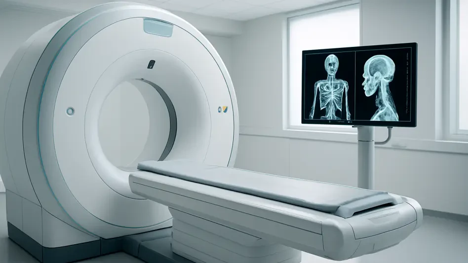
The arrival of photon-counting detector computed tomography in 2026 marked a revolutionary moment for the technical precision of pediatric death investigations. This advanced hardware offers a level of spatial resolution and tissue contrast that far surpasses conventional scanners, making it an indispensable tool for imaging the minute anatomical structures of fetuses and neonates. In the delicate context of pediatric cases, where a hairline rib fracture or a tiny intracranial hemorrhage can be the deciding factor in a diagnosis, the increased detail provided by this technology is a game-changer. It allows radiologists to visualize internal structures with a clarity that was previously only possible through high-magnification physical pathology. By capturing more detailed information from every photon that passes through the body, these scanners reduce image noise and improve the visualization of subtle pathologies. This leap in imaging quality has further narrowed the gap between digital investigation and dissection.
This technological evolution has been mirrored by a refinement in the clinical philosophy governing how such hardware is utilized within hospital settings. The focus has shifted from merely having the equipment to optimizing the entire workflow to serve the specific needs of postmortem subjects. Unlike living patients, the deceased present unique challenges such as tissue cooling and the cessation of blood flow, which affect image quality. Between 2024 and 2026, specialized protocols were refined to account for these physiological changes, ensuring that the data recovered from a scan is as accurate as possible. This meticulous approach has helped move postmortem imaging from a “last resort” for families who refuse autopsy to a preferred first-line diagnostic tool in many leading academic centers. The ability to perform a complete “virtual autopsy” within minutes has also streamlined the investigative process, allowing for faster release of the body to the family for funeral rites.
Global Adoption: Addressing Funding and Accessibility Gaps
While the scientific and technical achievements of the decade are impressive, the global adoption of postmortem imaging remains geographically uneven due to persistent economic and institutional barriers. In many regions, particularly across North America, the integration of these services has faced hurdles related to funding models and institutional inertia. Postmortem imaging often exists in a budgetary “grey zone” where it is not classified as a standard clinical diagnostic for a living patient nor always recognized as a reimbursable forensic expense. Consequently, the availability of these advanced services is frequently limited to well-resourced academic institutions, creating a disparity in the quality of death investigation accessible to families based on their location. Addressing these financial challenges requires a systemic rethink of how hospitals and legal systems value the information gained from postmortem examinations. Sustainable funding is required to reach more families.
To overcome these barriers and promote a standardized global approach, the international medical community recently reached a consensus through a major multi-society statement issued in 2026. This document, endorsed by radiological bodies from Europe, North America, Latin America, and Asia, establishes a global benchmark for the implementation of postmortem imaging services. It serves as a powerful advocacy tool for health administrators and government officials, signaling that these techniques are no longer peripheral experiments but are a recognized standard of care. By presenting a unified front, the global medical community has provided the necessary leverage to demand better funding and support for postmortem services worldwide. This international coalition also facilitates the sharing of expertise and the development of educational programs, ensuring that radiologists in emerging markets can be trained in the latest techniques and contribute to the growing global database of pediatric cases.
A New Methodology: Integrating Radiology with Traditional Pathology
The successful integration of imaging into the forensic toolkit transformed the landscape of pediatric medicine by establishing a more compassionate and versatile approach to death investigation. This evolution proved that the dead can indeed teach the living when modern technology is applied with sensitivity and scientific rigor. Moving forward, the focus shifted toward making these services a permanent part of the multidisciplinary team, where radiologists and pathologists worked in tandem to provide the most comprehensive answers possible. Actionable next steps for health systems included the formalization of training fellowships in postmortem radiology to ensure a steady pipeline of experts and the integration of imaging data into national health registries. These measures were designed to maximize the long-term utility of the information gathered, directly informing public health policies and neonatal care standards to improve outcomes for all future patients in the medical system.
Looking ahead, the commitment to standardized and culturally sensitive systems remained the cornerstone of modern pediatric medicine. The medical community recognized that the lessons learned from every loss had to be preserved to protect future generations, fulfilling the timeless promise that the quietest patients could still speak through the power of science. By treating imaging as a standard component of the diagnostic process, clinicians ensured that no vital medical data was lost due to the refusal of invasive procedures. The ongoing development of remote reporting networks allowed specialized expertise to reach underserved areas, further democratizing access to high-quality investigations. Ultimately, the transformation of the last decade solidified a new standard of care, ensuring that every investigation was conducted with the highest degree of accuracy, respect, and compassion for the families involved, ensuring that the legacy of each child contributed to the safety of others.