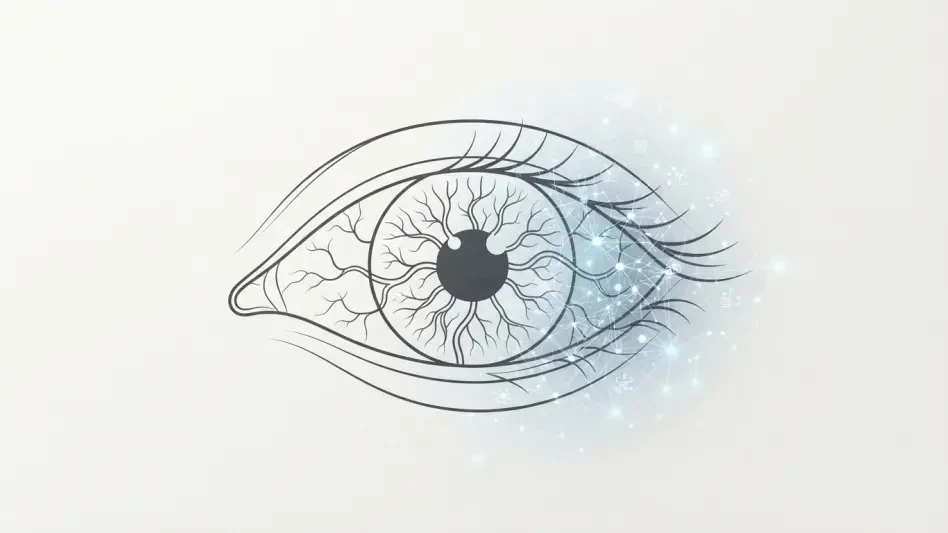

The primary technical challenge in retinal screening lies in the quality of data collected in real-world clinical settings where pupil dilation is often inconvenient or uncomfortable. This reality has long hindered the widespread adoption of ocular diagnostics for systemic diseases, yet the field of oculomics is now pushing past these constraints to transform the human eye into a sophisticated diagnostic portal. Researchers at Arizona State University, supported by a five-year, $3.2 million grant from the National Institutes of Health, are pioneering a method to detect Alzheimer’s disease through high-resolution retinal imaging. By focusing on the eye as a non-invasive biological window, this interdisciplinary effort aims to identify neurodegenerative markers years before the first signs of memory loss appear. The study represents a shift from reactive medicine to proactive screening, utilizing advanced artificial intelligence to analyze the complex microvasculature and neural structures of the retina, which serve as indicators of cognitive health.
Bridging the Gap: The Biological Link Between Eye and Brain
The fundamental connection between the retina and the central nervous system is more than just proximity; the eye is essentially a displaced piece of the brain that remains accessible to external observation. During embryonic development, the retina emerges from the same neural tube tissues that form the brain, leading to shared anatomical and physiological characteristics. Because of this shared origin, the vascular networks and nerve fiber layers in the back of the eye often mirror the condition of those within the skull. When neurodegenerative processes begin to take hold, the changes are not confined to the brain’s hidden regions but are instead reflected in the retinal tissue. This biological mirror allows clinicians to observe the health of the nervous system without resorting to invasive surgeries or expensive imaging technologies, providing a clear path toward understanding how systemic diseases progress at a microscopic level throughout the human body.
Identifying the specific hallmarks of Alzheimer’s, such as the accumulation of amyloid-beta plaques and tau protein tangles, has traditionally required specialized brain scans or uncomfortable lumbar punctures. However, recent advancements in ocular research indicate that these same proteins can manifest in the retinal layers, accompanied by a measurable thinning of the retinal nerve fiber layer. These subtle physiological shifts often precede any observable cognitive decline, offering a critical window for intervention. By using high-resolution imaging to map these changes, scientists are creating a topographical record of neurodegeneration that matches the atrophy seen in the cerebral cortex. This approach eliminates many of the barriers associated with traditional diagnostics, as the retina can be imaged repeatedly over time to track disease progression with high precision. Such continuous monitoring is vital for understanding early stages where the most significant opportunities for treatment are found.
Overcoming Technical Barriers: The Role of Generative AI
The practical application of retinal diagnostics frequently encounters the hurdle of inconsistent image quality, as many patients find the process of pupil dilation to be an intrusive part of a standard eye examination. Without dilation, photographs of the retina are often obscured by poor lighting, motion blur, or inadequate resolution, making them unsuitable for detailed medical analysis. To address this limitation, the research team is deploying generative artificial intelligence to enhance and reconstruct these low-fidelity images into high-quality data points. Unlike standard image filters, these AI models are designed to identify and clarify the underlying biological structures without introducing artificial distortions or misleading artifacts. This image rescue process allows researchers to utilize data that would typically be discarded, significantly increasing the pool of available information for diagnostic screening. By refining the visual output from standard clinical cameras, the technology makes it possible to conduct high-level neurological assessments.
Beyond simple image enhancement, the project utilizes complex deep learning algorithms to analyze the intricate patterns of the retinal microvasculature. These models are trained to detect minute changes in the density and branching of blood vessels that may signal the onset of Alzheimer’s disease. By processing thousands of high-resolution images, the AI can recognize pathological signatures that are far too subtle for the human eye to perceive during a routine clinical review. This automated analysis provides an objective and standardized method for assessing risk, reducing the likelihood of human error and ensuring that every patient receives a consistent level of scrutiny. The integration of generative AI into this workflow serves as a bridge between the limitations of current hardware and the requirements of modern precision medicine. As the algorithms continue to learn from diverse datasets, their ability to differentiate between normal aging and early-stage neurodegeneration becomes increasingly refined.
Enhancing Accuracy: Multimodal Data and Risk Assessment
One of the most significant challenges in current Alzheimer’s diagnostics is the existence of a gray zone where patients show ambiguous results on standard tests, such as blood-based biomarker evaluations. While these blood tests can categorize some individuals as clearly high-risk or low-risk, a substantial portion of the population falls into an indeterminate middle category that leaves clinicians without clear direction. The AI system addresses this problem by incorporating retinal data as an additional, independent layer of evidence, creating a multimodal diagnostic framework. By combining the visual insights from the eye with genetic information, age, and sex, the technology produces a more comprehensive and individualized risk profile. This layering of information allows for a much more nuanced interpretation of a patient’s health status, providing the necessary context to resolve clinical ambiguity. When retinal patterns are found in conjunction with specific blood markers, the confidence of a diagnosis increases.
This multimodal strategy represents a departure from the traditional diagnostic model that focuses on a single marker in isolation. Instead, the AI-driven approach views neurodegeneration as a complex, multifaceted process that requires a holistic view of the patient’s biological data. By integrating diverse datasets, the system can identify unique risk signatures that might be missed if the retina or blood markers were analyzed separately. This predictive capability is particularly valuable for identifying individuals who are at risk of rapid cognitive decline, allowing for the early implementation of personalized lifestyle interventions or therapeutic regimens. The goal is to move toward a model of precision health where every piece of data, from the microscopic vessels in the eye to the proteins in the blood, contributes to a unified understanding of brain health. This comprehensive view not only improves the accuracy of early detection but also provides a roadmap for monitoring how different patients respond to various preventative measures.
Expanding Access: Institutional Collaboration and Rural Outreach
The success of this diagnostic initiative is underpinned by a robust network of institutional partnerships that bridge the gap between academic research and clinical practice. Collaborators from the Mayo Clinic provide essential neuro-ophthalmological expertise, ensuring that the AI models are grounded in the realities of medical science and patient care. Meanwhile, partnerships with the Banner Alzheimer’s Institute and the Translational Genomics Research Institute offer access to vast and diverse patient datasets, which are crucial for validating the technology across different populations. These collaborations enable the research team to refine their algorithms using real-world data from individuals with varying health backgrounds and genetic profiles. This collective approach ensures that the resulting diagnostic tools are not only technically sound but also clinically relevant and broadly applicable. By pooling the resources of leading medical institutions, the project is able to accelerate the development of a screening system.
A core objective of this collaborative effort is to democratize access to advanced neurological screening, particularly for individuals living in rural or underserved regions. In many areas of Arizona and the broader United States, access to specialized memory care centers is limited by geography and the high cost of sophisticated diagnostic imaging. The development of an inexpensive and portable retinal screening tool offers a solution to this disparity, potentially allowing local optometrists and general practitioners to conduct early-stage Alzheimer’s evaluations. By utilizing mobile laboratories and standard clinical equipment enhanced by AI, the technology can reach populations that have historically been excluded from early intervention programs. This focus on accessibility is a critical component of the mission, as it ensures that the benefits of cutting-edge research are available regardless of location. Making early detection a routine part of community healthcare could significantly reduce the long-term burden on the medical system.
Strategic Importance: Preclinical Detection and Prevention
Shifting the focus of Alzheimer’s diagnostics to the preclinical stage is widely regarded as the most important advancement in the field, as it allows for the identification of the disease before any irreversible damage occurs. Extensive research has shown that by the time a patient begins to exhibit symptoms like memory loss or cognitive confusion, the underlying neurodegenerative process has already been active for years, if not decades. At this point, many therapeutic interventions are less effective because the neural pathways they are designed to protect have already suffered significant atrophy. The use of AI-enhanced retinal imaging provides a scalable and non-invasive way to detect these early warning signs during a window of time when preventative strategies are most likely to succeed. This earlier detection timeline is also essential for the progress of clinical trials, as it enables researchers to enroll participants who are still cognitively healthy but show biological markers of high risk.
The recent progress in ocular-based diagnostics established a new framework for managing brain health through the lens of preventative care. Researchers and clinicians successfully demonstrated that the integration of artificial intelligence with retinal imaging could provide a reliable and accessible method for early Alzheimer’s screening. By moving away from the invasive and expensive protocols of the past, the medical community took a significant step toward making neurological evaluations as routine as a standard physical examination. The project highlighted the necessity of interdisciplinary cooperation, showing how the combination of engineering, genomics, and clinical medicine could solve complex challenges in modern healthcare. Ultimately, the work focused on providing actionable insights that empowered individuals to take control of their neurological well-being long before symptoms manifested. The shift toward a predictive medical model represented a fundamental change in the global strategy against neurodegeneration, emphasizing vascular and neural health.