
A 54-year-old traveler returning to Bogotá, Colombia, recently presented with an itchy, painful nodule that appeared to be a standard skin infection but was actually something far more biological. While a tiny central pore might suggest the presence of a parasite, many clinicians mistakenly default to a diagnosis of cellulitis or a sebaceous cyst. This diagnostic pitfall often occurs because the initial symptoms of cutaneous myiasis—a localized infestation by fly larvae—mimic common dermatological conditions like furuncles or abscesses. The human botfly, known scientifically as Dermatobia hominis, relies on a sophisticated life cycle where eggs are transported to human hosts via blood-sucking vectors like mosquitoes. Once the larva burrows into the subcutaneous tissue, it creates a dome-shaped, erythematous lesion that can easily be misinterpreted as a bacterial problem. In the absence of specialized imaging, patients often receive unnecessary antibiotics or undergo invasive incisions that risk rupturing the parasite, leading to severe inflammatory complications. By shifting the focus to high-frequency ultrasound, medical professionals can achieve a level of diagnostic certainty that was previously unattainable in routine clinical settings, ensuring that patients receive appropriate, targeted care without the trauma of misdirected surgical interventions.
Identifying the Parasite Through High-Frequency Imaging
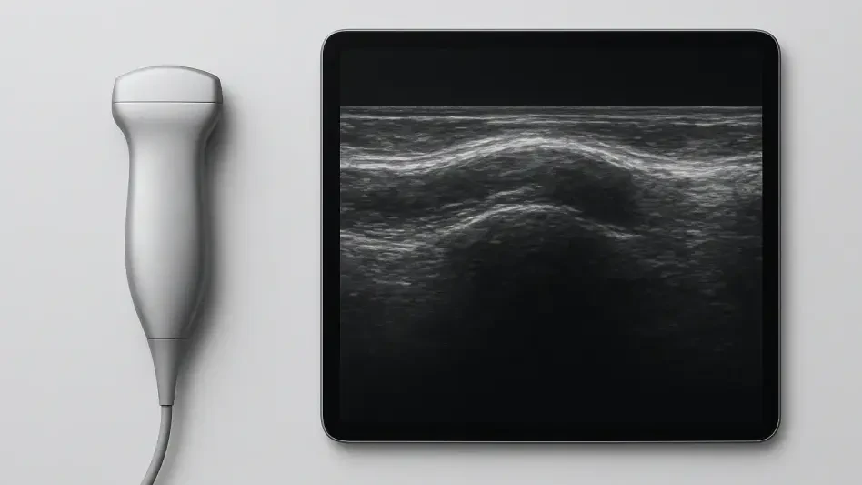
The integration of 13-MHz high-frequency linear transducers into the diagnostic workflow represents a major leap forward in managing soft-tissue parasitic infections. These specialized probes provide the high spatial resolution required to visualize structures just millimeters beneath the skin surface, making them ideal for identifying the human botfly larva. Unlike traditional low-frequency ultrasound, which is better suited for deep abdominal organs, high-frequency imaging clarifies the distinction between a liquid-filled abscess and a solid biological entity. On a standard gray-scale or B-mode image, the larva appears as an echogenic structure, meaning it reflects sound waves more intensely than the surrounding subcutaneous fat. This visual differentiation is critical; whereas an abscess appears dark or hypoechoic due to its fluid content, the presence of an organized, reflective mass within the fat layer immediately suggests a foreign or parasitic origin. This allows the radiologist to move beyond the surface-level appearance of the lesion and peer into the architecture of the subcutaneous tissue, identifying the physical presence of the organism long before any invasive measures are considered.
A specific radiological hallmark of myiasis is the presence of posterior acoustic shadowing, a phenomenon that occurs when a dense structure blocks or absorbs the ultrasound beam. In the case of the botfly larva, its tough chitinous exoskeleton acts as a barrier, preventing sound waves from penetrating deeper and creating a distinct dark area or shadow directly beneath the organism. This finding is highly significant because it confirms that the lesion is not a simple collection of pus or serum, which would allow the sound waves to pass through relatively unimpeded. While other foreign bodies, such as wooden splinters or glass fragments, can also cause shadowing, the characteristic oval shape and localized inflammatory response associated with the larva provide a specific visual signature. When a clinician observes this combination of an echogenic mass and a clear acoustic shadow, they can effectively rule out common skin infections and focus on the likelihood of a parasitic infestation. This precision reduces the reliance on clinical guesswork and helps prevent the accidental rupture of the larva during a standard incision and drainage procedure, which is a common error in non-endemic medical environments.
Using Doppler and Dynamic Motion for Confirmation
Color Doppler ultrasonography adds a dynamic layer to the diagnostic process by mapping the movement of blood in the tissues surrounding the suspected parasite. In cases of cutaneous myiasis, the host’s immune system responds aggressively to the presence of the larva and its metabolic waste, leading to a state of intense local hyperemia. On the ultrasound screen, this appears as a vibrant ring of color—representing increased blood flow—encircling the echogenic mass. This inflammatory signature is vital for distinguishing a parasitic infestation from a non-living foreign body or a stagnant cyst. Furthermore, Doppler imaging allows medical professionals to differentiate the larva from vascular tumors or other growths that might otherwise appear similar on a basic gray-scale scan. While a tumor would typically show blood flow signals directly within the mass itself, a fly larva is an independent organism and does not share the host’s circulatory system. Therefore, the absence of internal vascular signals, combined with the intense peripheral blood flow, provides a clear roadmap for the clinician, confirming the presence of an active, inflammatory biological process without the need for biopsy.
The most compelling evidence for a living parasite is captured through real-time dynamic assessment, where the actual movements of the larva are observed on the ultrasound monitor. Radiologists can witness spontaneous, rhythmic contractions—resembling peristalsis—as the larva shifts within its subcutaneous burrow. This wriggling motion is considered pathognomonic, meaning it is an unmistakable indicator of a living organism and is not found in any other skin condition that mimics myiasis. Observing these contractions for as little as thirty to sixty seconds can provide the smoking gun required for a definitive diagnosis. It transforms the patient’s subjective report of a crawling sensation into a visible, objective reality. This real-time visualization is particularly powerful in a clinical setting, as it allows the medical team to observe how the larva interacts with the surrounding tissue and how it responds to external pressure from the ultrasound transducer. By documenting these active muscular movements, physicians can move forward with extraction procedures with absolute confidence, knowing that they are dealing with a live parasite rather than a dormant cyst or a bacterial infection.
Strategic Advances in Clinical Management
Confirming the location and orientation of the larva through advanced imaging allows for a highly strategic approach to medical management and extraction. Traditional treatment often involved blind maneuvers that risked damaging the larva, but ultrasound-guided planning enables a much gentler and more effective intervention. Clinicians can accurately measure the depth of the parasite and identify the position of its breathing pore, which is essential for successful occlusion therapy. This method involves covering the central pore with substances like petrolatum, beeswax, or surgical tape to deprive the larva of oxygen. As the parasite moves toward the surface to find air, its progress can be monitored in real-time, allowing medical professionals to grasp it with forceps at the optimal moment. This targeted approach minimizes trauma to the surrounding skin and reduces the risk of secondary infections that often follow more aggressive surgical attempts. By utilizing the insights gained from high-frequency scans, the healthcare team can ensure that the extraction is performed cleanly, maintaining the integrity of the parasite’s body and preventing the release of allergenic proteins into the host’s system.
The utility of Doppler ultrasound extends beyond the initial diagnosis and into the post-extraction phase of care, serving as a critical tool for procedural verification. After the larva has been removed, a secondary scan can be performed to ensure that the subcutaneous cavity is entirely clear of any biological remnants. This step is vital because even small fragments of the larva’s chitinous exoskeleton can trigger a chronic foreign-body granuloma, leading to persistent inflammation and the need for further surgical intervention. By confirming the empty cavity status through ultrasound, doctors can provide patients with immediate peace of mind and prevent long-term complications. Additionally, the imaging can track the resolution of the surrounding hyperemia, providing a visual record of the healing process as the inflammatory response subsides. This comprehensive approach to management—from the first diagnostic scan to the final verification—represents a significant improvement over traditional methods that relied heavily on visual inspection and patient feedback. It ensures a higher standard of clinical accuracy and a more streamlined recovery for individuals suffering from this distressing condition.
Global Impact on Travel and Tropical Medicine
In the current medical landscape of 2026, the rise in international travel to tropical and subtropical regions has made the recognition of exotic conditions like myiasis increasingly important for clinicians worldwide. As more individuals visit endemic areas in Central and South America or sub-Saharan Africa, the likelihood of patients returning with parasitic infestations continues to grow. This shift necessitates a broader adoption of specialized diagnostic protocols in temperate climates, where such cases were once considered rare. The accessibility of high-frequency ultrasound equipment in modern emergency departments and dermatology clinics provides a practical solution to this challenge, allowing for rapid, point-of-care diagnostics. Instead of waiting for laboratory results or specialist consultations, frontline physicians can use handheld or console-based ultrasound systems to provide an immediate answer. This technological democratization ensures that patients receive the correct diagnosis during their initial visit, preventing the cycle of misdiagnosis and ineffective antibiotic use that has historically characterized the management of these cases in non-endemic urban centers.
The successful application of Doppler and dynamic imaging in recent clinical cases demonstrated that the most effective tool for managing subcutaneous myiasis was already available in many standard medical facilities. Clinicians learned that by integrating travel history with high-frequency scans, they could avoid the high costs and physical toll of misdirected treatments. Moving forward, the medical community should prioritize the development of standardized ultrasound training for the detection of parasitic organisms, ensuring that radiologists and emergency physicians are familiar with the wriggling signature of living larvae. This case established that non-invasive visualization not only improved diagnostic speed but also enhanced the safety of the extraction process by providing a clear anatomical map for intervention. Future efforts should focus on creating accessible digital libraries of dynamic ultrasound clips to help clinicians in 2026 and beyond recognize these patterns more quickly. Ultimately, the transition toward point-of-care imaging simplified a complex tropical disease into a manageable clinical task, proving that the synergy between traditional parasitology and modern technology is essential for addressing the health challenges of an increasingly interconnected global population.