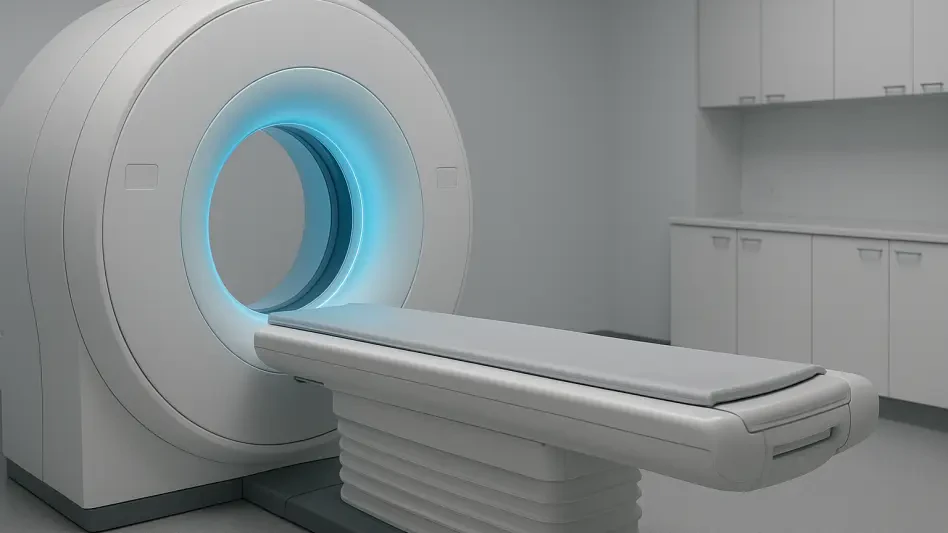

Accurate radiation dose calculations are often hindered by the truncation-induced density shifts that occur when peripheral structures like shoulders are excluded. In the precision-driven world of oncology and orthopedic surgery, even a minor discrepancy in anatomical representation can lead to significant variations in treatment efficacy or diagnostic accuracy. Cone-beam computed tomography, or CBCT, has revolutionized point-of-care imaging by providing detailed three-dimensional insights within compact clinical settings. However, the physical constraints of detector panels often result in incomplete data capture, leaving clinicians to work with images that are marred by severe peripheral distortions. Recent breakthroughs in artificial intelligence are now offering a way to bypass these hardware limitations by synthesizing the missing anatomical information. By integrating advanced generative models into the reconstruction pipeline, researchers have found a method to restore the integrity of these scans, ensuring that the final output remains a faithful representation of the patient’s true physical state.
The Technical Challenge: Persistent Image Truncation
The mechanics of CBCT differ significantly from traditional multi-detector CT scans, as they use a cone-shaped X-ray beam and a flat detector to capture data in a single rotation. While this setup allows for rapid imaging and high spatial resolution, the human body is frequently larger than the active area of the detector panel. When peripheral structures like the shoulders or the edges of the skull are not captured, the resulting “truncated projections” create significant hurdles for the standard reconstruction algorithms. This phenomenon is not merely an aesthetic issue; it leads to a fundamental loss of information that the system needs to calculate the internal density of the scanned volume. Without this data, the math behind the image creation fails to account for the attenuation of X-rays passing through the missing tissue, leading to a cascade of errors that degrade the final 3D volume. This physical constraint has long been a limiting factor for expanding CBCT use into larger anatomical regions.
Understanding CBCT Mechanics and Artifacts
The Filtered Backprojection (FBP) algorithm, which is the standard for building 3D images from raw X-ray data, requires a complete dataset to function correctly. When data points are missing due to truncation, the algorithm generates artifacts that appear as artificial bright spots, distorted tissue intensities, or distracting streaks across the image. These errors can hide lesions and interfere with the precise density measurements necessary for planning radiation therapy or identifying diseased tissue.
Furthermore, these artifacts often manifest as a characteristic “cupping” effect, where the edges of the scan appear unnaturally bright, masking the true contrast of the peripheral anatomy. In clinical practice, this means that a tumor located near the edge of the field of view might be obscured or its margins misidentified. The resulting inaccuracy forces clinicians to rely on additional, often more invasive or expensive, imaging modalities to confirm findings that should have been clear in the initial scan.
Limitations of Traditional Remediation Methods
Historically, clinicians have used basic algorithmic “guesses” to fill in these missing gaps, often relying on software toolkits to pad the edges of the projection data. This traditional padding usually involves mirroring existing pixels at the detector’s boundary or applying a smooth decay function to bridge the gap toward zero. While these methods can reduce the most obvious visual distortions, they do not incorporate real anatomical information, often leaving the edges of the scan looking blurry or structurally inaccurate.
The failure of these geometric methods is most apparent in complex areas where tissue density changes rapidly. Because simple padding treats the missing space as a uniform extension of the last known pixel, it cannot account for the intricate variations of bones, muscles, and air pockets. Consequently, the quantitative reliability of the scan remains low. This lack of fidelity prevents the use of these images for automated diagnostic systems and precise surgical navigation, which require a near-perfect representation of the patient’s internal structure.
The Generative Revolution: Moving Beyond Traditional AI
To address the failures of traditional padding, research teams have turned to generative artificial intelligence, specifically exploring the potential of diffusion models to outperform existing neural networks. For several years, Generative Adversarial Networks (GANs) were the primary focus for image completion tasks. However, GANs are prone to “hallucinating” realistic-looking details that do not actually exist in the patient, a dangerous trait in a medical setting. These hallucinations could lead to a misdiagnosis if a physician mistakes an AI-generated artifact for a real pathological feature. The inherent instability of GAN training often results in images that look convincing but lack the mathematical grounding required for clinical safety. This has led to a search for more stable generative architectures that prioritize structural accuracy over mere visual plausibility, ensuring that every synthesized pixel is a reflection of biological reality rather than a statistical guess by the model.
Moving Beyond Hallucinations in AI
Conditional generative diffusion models offer a more stable alternative by using a controlled denoising process to reconstruct missing data. The model is trained to gradually remove noise from an image while being “conditioned” on the existing, non-truncated parts of the scan. This process forces the AI to fill in the missing segments in a way that is statistically and mathematically tied to the actual measured data. Unlike GANs, which compete to fool a discriminator, diffusion models focus on learning the underlying data distribution.
This approach ensures that the generated portions of the image are structurally consistent with the patient’s unique anatomy. By prioritizing fidelity, the diffusion model provides a reliable dataset for the reconstruction algorithm to process. This transition from adversarial competition to iterative denoising has proven to be a turning point in medical AI, offering a path toward synthetic data that clinicians can trust for diagnostic purposes. The resulting images are virtually indistinguishable from full-field-of-view scans in terms of their anatomical accuracy.
Reliability of Denoising Processes
The denoising mechanism within these models allows for a high degree of precision when predicting the density of missing tissues. By analyzing the global context of the captured X-ray projections, the AI can infer the presence of structures like the humeral head or the outer table of the skull with remarkable accuracy. This is achieved by reversing a diffusion process that has learned the complex spatial relationships within human anatomy. The result is a seamless integration of real and synthesized data that maintains the integrity of the Hounsfield units.
Moreover, the stability of this method makes it suitable for diverse patient populations, as the conditioning ensures the AI adapts to specific anatomical variations rather than applying a generic template. This robustness is critical for maintaining consistent performance across different scanner models and clinical settings. As these models matured, they provided a level of detail that traditional methods could not reach, effectively turning a limited detector into a virtual wide-field sensor through the power of mathematical inference.
Quantitative Validation: Proving the Power of AI
The effectiveness of the diffusion-based approach is supported by rigorous performance metrics, specifically Peak Signal-to-Noise Ratio (PSNR) and the Structural Similarity Index (SSIM). At the raw projection stage, diffusion models showed a massive 12-decibel improvement in PSNR over traditional padding methods. In the field of signal processing, such a leap represents a significant reduction in the error energy of the image, leading to much clearer 3D volumes. The SSIM scores, which measure how well the AI preserves the shapes and textures of the original anatomy, also showed substantial gains. These numbers translate directly to clinical value, as they indicate that the reconstructed images are not only visually cleaner but also maintain the structural integrity necessary for medical analysis. This quantitative proof has been essential in convincing the medical community that AI-driven data synthesis is a viable and safe alternative to larger hardware.
Measuring Performance and Image Fidelity
Beyond the global metrics, the clinical benefit was most evident in the improved visibility of low-contrast structures. In oncology, the ability to distinguish between a tumor and healthy tissue often relies on subtle gray-scale differences that truncation artifacts usually destroy. By preserving these nuances, the diffusion model allowed for more accurate diagnoses and better-informed treatment plans. This was particularly noticeable in soft-tissue imaging, where high-resolution detail is paramount for surgical success.
The removal of peripheral streaks and haze also improved the performance of automated segmentation tools. These software programs, which are used to outline organs for radiation therapy, often struggle with the noise introduced by truncation. With the AI-enhanced scans, the segmentation became more reliable, reducing the need for manual corrections by medical physicists. This improvement in image fidelity served as a bridge between raw data collection and the advanced diagnostic software that defines modern clinical practice.
Broader Implications for Global Healthcare
The implications of this technology reach across various medical disciplines, from dentistry to interventional radiology. Because this is a software-based solution, it was integrated into existing CBCT machines via simple updates, avoiding the need for expensive hardware overhauls. This provided a cost-effective way to enhance diagnostic capabilities worldwide, ensuring that even smaller clinics with older equipment could produce high-quality, artifact-free images. The digital nature of the enhancement allowed for rapid scaling across different healthcare systems.
Ultimately, the fusion of deep learning and classical physics established a new standard for image fidelity in 2026. This research suggested that hardware limits would no longer define the quality of clinical vision, as generative AI continued to act as a bridge between physical sensors and mathematical needs. By shifting the focus toward intelligent data restoration, the medical community took a significant step toward universal access to high-precision imaging, ensuring that patient safety and diagnostic accuracy remained at the forefront of technological progress.