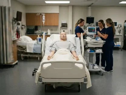

Foundational anatomy instruction is being redefined by tools that allow students to visualize the movement of muscles and nerves in a living subject. This shift reflects a broader modernization within medical schools where the static nature of cadaveric dissection is increasingly supplemented by dynamic, real-time diagnostic imaging. A comprehensive study published in BMC Medical Education by R. Miranda-Krause and her colleagues recently investigated this pedagogical evolution by examining how ultrasound technology impacts the learning experience of first-year medical students. By focusing on the topographical anatomy of the upper limb, the researchers sought to determine if observing live physiological structures provides a measurable advantage over traditional laboratory methods. The objective was to bridge the persistent gap between theoretical anatomical knowledge and the practical, clinical application required in modern healthcare environments, where point-of-care ultrasound has become an essential diagnostic standard.
Examining the Study Design and Methodology
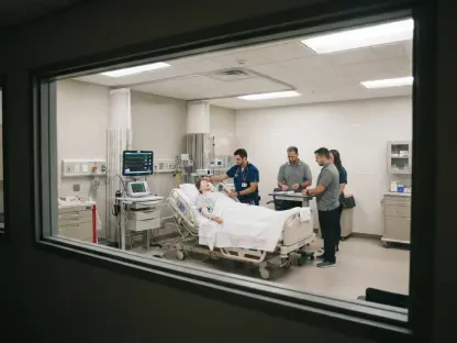
The research utilized a robust cohort of 79 first-year medical students to explore the efficacy of modern imaging versus traditional teaching tools. To facilitate the investigation, participants were divided into an intervention group consisting of 42 students and a control group of 37 students. This division was based on pre-existing laboratory sections to maintain logistical efficiency, which defined the study as a quasi-experimental design. Throughout the curriculum sessions, students in the intervention arm rotated through structured learning stations that integrated real-time ultrasound imaging of a living subject. This allowed them to observe the immediate contraction of muscles and the pulsation of vascular structures, contrasting sharply with the static models used by their peers. Meanwhile, the control group relied exclusively on traditional materials including anatomical models, detailed atlases, and cadaveric specimens, ensuring that all participants covered the same anatomical concepts.
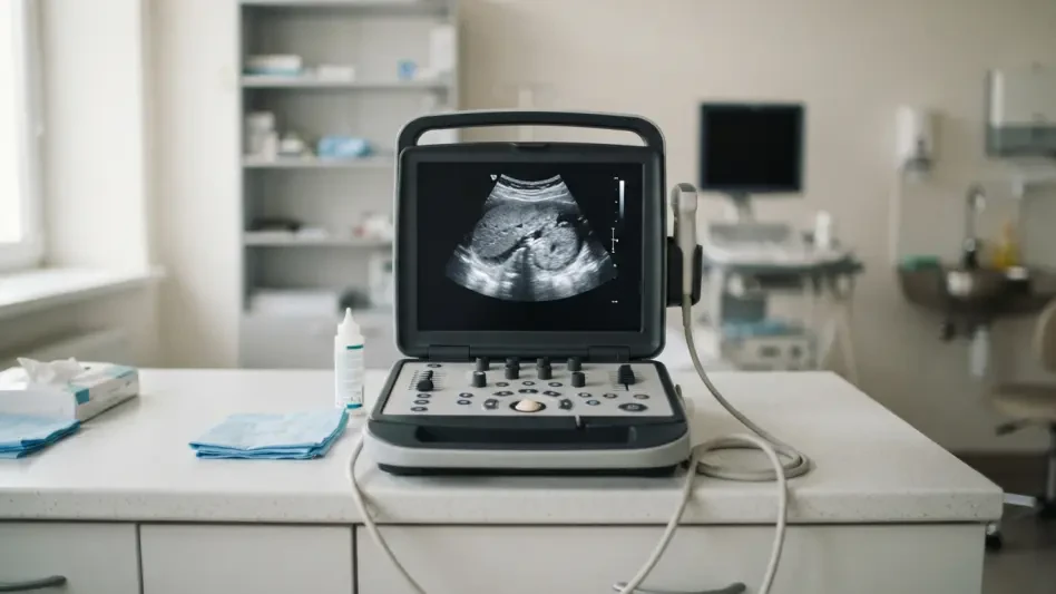
The educational intervention specifically targeted the regional anatomy of the upper limb, a complex area where spatial relationships are critical for clinical success. In the experimental condition, the laboratory session was organized into six distinct learning stations, three of which were enhanced with high-definition ultrasound equipment. These specialized stations required students to identify and track muscular, vascular, and neural structures as they functioned in real time. In contrast, the control group utilized high-quality cadaveric materials and physical models to achieve the same instructional objectives without the aid of dynamic imaging. To evaluate the impact of these different approaches, the researchers implemented a 15-item multiple-choice assessment. This evaluation was administered as a pretest to establish a baseline and as a posttest one week after the laboratory session, providing a clear window into how each method influenced short-term knowledge retention and anatomical comprehension.
Analyzing Quantitative Learning Outcomes
Initial findings from the quantitative data suggested that the technological intervention did not necessarily result in a superior grasp of factual knowledge. At the start of the study, both groups demonstrated nearly identical baseline scores, with a pretest p-value of 0.885 indicating no significant difference in prior knowledge. Following the instructional sessions, both the ultrasound-enhanced group and the traditional control group showed statistically significant improvements in their posttest scores. This outcome confirmed that both teaching modalities were highly effective in conveying the required anatomical information. However, the anticipated “silver bullet” effect of ultrasound was notably absent in the final metrics. The posttest performance revealed no significant statistical difference between the two cohorts, with the average score gap measuring a mere 0.69 points, suggesting that while technology is engaging, it may not immediately translate into higher test scores.
Statistical modeling via a 2 × 2 mixed-model analysis of variance further reinforced the conclusion that ultrasound did not provide a unique academic advantage in this context. While the intervention group displayed a slightly higher within-group effect size of 0.78 compared to the control group’s 0.59, the between-group difference remained minimal at an effect size of 0.23. One of the most intriguing patterns observed by the researchers was an inverse relationship between initial performance and subsequent learning gains. Students who began with the lowest pretest scores showed the most substantial improvement after the laboratory sessions, regardless of which group they were in. This indicates that the foundational curriculum was exceptionally beneficial for those starting with less prior knowledge. The medium of instruction—whether a high-tech ultrasound probe or a classic anatomical atlas—seemed secondary to the quality of the structured instructional content provided to the students.
Assessing Student Perceptions and Motivation
Although the quantitative data failed to distinguish one method as academically superior, the qualitative insights gathered from the students told a very different story. Through Likert-scale questionnaires and semi-structured interviews, participants expressed an overwhelming preference for the inclusion of ultrasound in their studies. They described the experience of seeing “living anatomy” as transformative, noting that it made abstract concepts significantly more tangible and easier to grasp. This subjective feedback highlighted a substantial increase in student motivation and classroom engagement when imaging technology was present. Many students reported that the ability to see a nerve or vessel move in real time provided a sense of clarity that static cadavers simply could not match. This psychological boost is a critical factor in medical education, as high levels of learner engagement are often correlated with greater persistence and a more positive overall educational environment.
The perception of clinical relevance emerged as a dominant theme in the qualitative analysis of student feedback. By introducing a diagnostic tool that is widely used in modern hospitals, the curriculum helped students bridge the gap between their introductory basic science coursework and their future roles as practicing physicians. Participants noted that using ultrasound made their first-year studies feel more purposeful and directly applicable to the clinical environment they aspire to join. This early exposure to diagnostic imaging appears to play a vital role in professional identity formation, allowing students to see themselves as clinicians in training rather than just students of biology. While these benefits might not be reflected in a multiple-choice test conducted one week after a lab, they contribute to a deeper sense of professional readiness. The consensus among the cohort was that the technology provided a valuable window into the realities of modern medical practice and diagnostic procedure.
Future Perspectives and Educational Implications
It is important to consider several limiting factors that may have influenced the outcome of this specific investigation into ultrasound integration. The study focused exclusively on the topographical anatomy of the upper limb, which leaves open the question of whether the technology would be more effective in other regions of the body. For instance, areas such as the abdomen or the cardiovascular system involve significantly more dynamic movement, where the real-time capabilities of ultrasound might offer a more distinct advantage over static models. Additionally, the exposure to ultrasound was only partial, as it was integrated into just half of the learning stations during the session. This limited exposure may have diluted the measurable impact of the technology on overall test scores. Furthermore, the assessment of knowledge occurred only one week after the intervention, leaving the long-term retention of these anatomical concepts and the durability of the learning gains largely unexamined.
The study ultimately suggested that while ultrasound did not serve as a shortcut to higher academic grades, its value as a pedagogical tool remained substantial. Educators determined that integrating dynamic imaging fostered a more interactive learning environment that successfully connected basic science to clinical reality. The research indicated that future curricula should balance traditional cadaveric dissection with modern imaging to provide a comprehensive view of human anatomy. It was concluded that the most effective approach involved using ultrasound not as a replacement for established methods, but as a supplementary layer that enhanced student engagement and professional motivation. Moving forward, academic institutions were encouraged to implement longitudinal studies to track how early exposure to these tools influenced diagnostic accuracy in clinical rotations. By prioritizing a blended learning model, medical schools ensured that students developed both a deep foundational knowledge and the practical skills necessary for modern medicine.