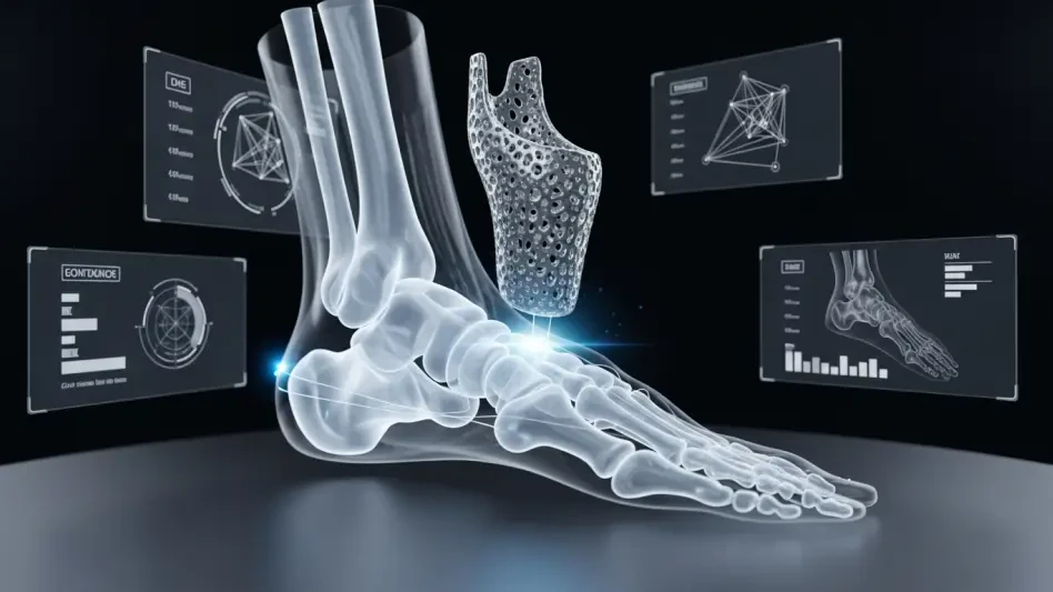

The intricate biological engineering of the human foot requires an astonishing level of coordination between bones, tendons, and ligaments, yet surgeons have historically repaired this complex system using generic, mass-produced metal plates. This reliance on off-the-shelf hardware often necessitated a compromising approach where the patient’s anatomy was altered to fit the implant. However, the rise of 3D printing technology is fundamentally changing the surgical landscape by prioritizing the patient’s unique skeletal structure. By shifting from a “one-size-fits-all” mentality to personalized, anatomy-driven solutions, the medical field is entering a period where precision is the baseline rather than the exception.
The End of Forced Fits: Orthopedic Reconstruction Evolution
For decades, orthopedic reconstruction followed a rigid protocol where surgeons selected from a limited inventory of standardized plates and screws. These components, while durable, were designed based on anatomical averages that failed to account for the subtle curves and densities of an individual’s bone structure. In practice, this meant that during highly complex procedures, medical teams frequently had to remove healthy bone or use excessive force to align hardware. This traditional “forced fit” approach often resulted in secondary issues, as the body struggled to adapt to metal that did not mirror its natural mechanics.
The introduction of 3D-printed titanium has effectively flipped this script, enabling the hardware to be designed around the patient. Instead of bending a plate to match a bone, engineers now use digital models to print implants that sit flush against the bone surface from the moment they are placed. This evolution reduces the mechanical stress on the surrounding tissue and significantly lowers the likelihood of hardware irritation. Consequently, the anatomical integrity of the foot and ankle is preserved, allowing for a more natural restoration of movement and function that was previously difficult to achieve with mass-produced alternatives.
Precision Standard: The New Benchmark for Lower Extremity Health
In the specialized field of foot and ankle surgery, the difference between a successful recovery and chronic pain is often a matter of a few millimeters. Minor misalignments in the lower extremities can trigger a domino effect, leading to altered gait patterns, premature joint wear, and eventual failure of the reconstruction. Standardized implants, while functional, lacked the specificity required to address complex deformities without creating these minor but significant discrepancies. As healthcare providers move toward value-based care, the demand for precision has become the primary driver for technological adoption.
The shift toward patient-specific solutions has established a new standard where “first-time-right” outcomes are the goal for every procedure. By utilizing custom-fabricated components, surgeons can ensure that every angle and attachment point is optimized for the patient’s specific weight-bearing needs. This level of accuracy is particularly vital in addressing long-term mobility, as it prevents the compensatory movements that often lead to secondary injuries in the knees or hips. High-precision hardware essentially serves as a preventive measure against the degenerative cycles typically associated with traditional orthopedic surgery.
Digital Architecture: Streamlining Complex Reconstructions
Modern surgical planning has moved beyond the two-dimensional constraints of traditional X-rays into a realm of sophisticated digital architecture. By integrating high-resolution CT imaging with advanced software, surgeons can visualize and manipulate a patient’s anatomy in a virtual environment long before the first incision is made. This digital-first workflow allows for the creation of patient-matched surgical guides that act as a roadmap during the operation. These tools dictate the exact placement of every screw and the precise angle of every cut, removing the guesswork that once characterized intraoperative adjustments.
This streamlined methodology is especially transformative for patients suffering from multiple co-existing pathologies or severe traumatic injuries. In the past, such cases often required multiple staged surgeries, resulting in months of recovery and increased risks of infection. Today, 3D printing allows medical teams to address several deformities in a single, unified procedure. By consolidating the reconstruction into one highly planned event, hospitals can reduce the time a patient spent under anesthesia. The result is a more efficient surgical process that benefits both the clinical team and the patient’s overall recovery timeline.
Economic Shift: Expert Perspectives on Clinical Progress
The transition to patient-specific technology is not merely a clinical improvement but a significant economic evolution within the healthcare system. Nancy Hairston, CEO of MedCAD, noted that the adoption of these solutions represents a fundamental shift in how hospitals evaluate the cost of care. While custom-printed implants might carry a higher initial price tag than bulk-purchased hardware, they offered substantial long-term savings by reducing the need for revision surgeries. Research suggests that the reduction in operating room time and the decrease in postoperative complications more than offset the upfront investment in personalized technology.
Expert consensus increasingly points to a future where standardized implants are reserved for only the most basic fractures. Healthcare institutions that resisted this shift toward anatomy-driven reconstruction found themselves at a disadvantage as patients began to expect higher standards of personalized care. The move toward patient-matched hardware aligned perfectly with the broader trend of precision medicine, where every intervention is tailored to the individual’s biological data. This economic logic made it clear that investing in better outcomes initially was far more sustainable than managing the costs of failed or suboptimal traditional surgeries.
Integration Framework: Adopting Patient-Matched Technology
The transition toward digital-first protocols marked a definitive departure from the trial-and-error methodologies of the past. Surgical teams that embraced advanced imaging protocols and collaborative manufacturing partnerships successfully established a new baseline for patient care. This shift not only streamlined the operative process but also ensured that the long-term durability of reconstructions became a predictable outcome rather than a statistical hope. As the industry looked ahead, the move to anatomy-driven restoration provided the necessary foundation for a more sustainable and effective healthcare model.
Surgeons who integrated these tools into their daily practice reported a significant decrease in intraoperative stress and a more intuitive surgical experience. By shifting the burden of planning from the operating room to the preoperative digital phase, clinics achieved a level of consistency that was previously unattainable. The partnership between medical professionals and technology experts facilitated a predictive model of care that prioritized the patient’s unique geometry. Ultimately, the adoption of 3D-printed solutions proved that when technology respected the body’s natural form, the results were both clinically superior and economically sound.