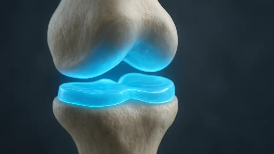

Automated segmentation tools allow for the early detection of joint degradation, potentially opening a window for intervention years before a patient experiences functional failure. Knee osteoarthritis is a pervasive health challenge that stands as a primary cause of chronic pain and disability in aging populations. The condition is defined by the progressive deterioration of articular cartilage, the vital tissue that ensures smooth joint movement. Traditionally, medical professionals have relied on subjective assessments to monitor this decay, but the most crucial changes occur at a microscopic level. Because this cartilage appears as an extremely thin, irregular ribbon on an MRI, identifying early-stage degradation requires a level of precision that is difficult to achieve consistently through manual human observation. The current gold standard for evaluating joint health is manual segmentation, a process where radiologists trace the boundaries of the cartilage pixel by pixel. This task is incredibly grueling, time-consuming, and highly susceptible to human error or variation between different observers. Even a tiny mistake in tracing can lead to significant inaccuracies in measuring cartilage volume or thickness. To solve this bottleneck, researchers have developed an automated deep learning approach designed to handle these complexities with expert-level accuracy, effectively removing the burden of manual labor from the diagnostic process and ensuring that measurements remain consistent across different clinical settings.
Technological Advancements in Musculoskeletal Imaging
Addressing Technical Limitations and Demographic Gaps
The development of this new AI model was largely motivated by an urgent need to overcome the inherent shortcomings of traditional convolutional neural networks, which have dominated medical imaging for years but often struggle to understand the relationship between distant parts of a complex image. While these networks are excellent at identifying local patterns, they frequently fail to grasp the “long-range dependencies” that are essential for accurate anatomical mapping. For thin and curvilinear structures like knee cartilage, the model must understand the broader anatomical context of the surrounding bone to define boundaries accurately. Without this holistic perspective, an algorithm might misidentify a shadow or a minor structural variation as a significant pathological change. By moving toward a Transformer-based architecture, the researchers have managed to integrate this necessary global context, allowing the system to verify the placement of cartilage against the sturdy, more easily identified landmarks of the femur and tibia. This systemic understanding ensures that the software can differentiate between healthy tissue and the early signs of thinning that characterize the onset of osteoarthritis.
Furthermore, the researchers aimed to fix a persistent “data gap” in the field of artificial intelligence, as most existing MRI datasets have historically been based on Western populations. This lack of diversity can lead to biases in diagnostic tools, as anatomical nuances and disease patterns often vary across different ethnicities and lifestyles. By creating a specialized dataset focused on Asian anatomy, the team has significantly improved the diagnostic reliability of AI tools for a more diverse global population. This effort is particularly relevant in 2026, as healthcare systems worldwide strive to implement more equitable diagnostic standards that do not sacrifice accuracy when applied to different demographic groups. Ensuring that a model can perform equally well on a variety of knee shapes and sizes is a critical step toward the universal adoption of AI in orthopedics. This focus on demographic inclusivity ensures that the benefits of early detection are not confined to a single region but can be shared by clinical practices around the globe, providing a more robust foundation for personalized medicine.
The OAMRI Dataset: A Research Benchmark
Central to this advancement is the creation and deployment of the OAMRI dataset, which includes nearly 900 high-resolution MRI slices meticulously curated to represent a wide spectrum of joint health. These images are T2-weighted, a specific type of imaging that is highly sensitive to both the physical structure of the cartilage and the subtle fluid changes that signal early-stage breakdown before visible thinning occurs. By utilizing T2-weighted scans, the researchers have provided the model with the richest possible data environment, allowing it to detect “biochemical” changes in the cartilage matrix. This level of detail is essential because, by the time physical erosion is visible on a standard X-ray or a lower-resolution scan, the opportunity for non-surgical intervention has often already passed. The dataset serves as a comprehensive map of the knee’s internal environment, capturing the interplay between bone density and tissue integrity in a way that allows the AI to learn the subtle hallmarks of degenerative disease across various stages of progression.
Expert orthopedic surgeons and senior radiologists spent countless hours meticulously labeling these images to identify the femur, the tibia, and their respective cartilages. This rigorous annotation process ensures that the AI’s training is grounded in the highest level of human expertise, effectively teaching the machine to see what a veteran specialist sees. By making this dataset open-access, the researchers have provided a vital resource that allows scientists worldwide to test, validate, and refine their own diagnostic algorithms. This commitment to open science is a cornerstone of current medical progress in 2026, as it encourages collaboration over competition. Providing a standardized benchmark means that different research teams can compare their results on a level playing field, accelerating the pace of innovation and ensuring that only the most reliable models make it into the clinical environment. The availability of such high-quality, expert-vetted data reduces the barrier to entry for smaller research institutions, fostering a more vibrant and competitive ecosystem for medical technology development.
Innovative Architecture and Model Performance
Utilizing Swin-Unet: Precise Segmentation
To process the intricate and often overlapping data points within the knee joint, the research team utilized the Swin-Unet architecture, which employs a “Transformer-based” approach rather than traditional pixel filtering. Unlike older models that look at an image through a small, fixed window, this method uses self-attention mechanisms to divide an MRI scan into small patches and analyze how they relate to one another across the entire image. This allows the model to “attend” to specific features, such as the contact point between the femur and tibia, while ignoring irrelevant background noise. The model’s “shifted window” technique is particularly effective in this regard, as it captures the fine details of thin cartilage edges while simultaneously maintaining a view of the entire joint’s mechanical alignment. This dual-focus capability is what sets the Transformer model apart, as it mimics the way a human expert might zoom in to look at a specific lesion while keeping the overall structure of the leg in mind to ensure anatomical consistency.
This technological shift is vital because the boundaries between cartilage and other soft tissues in the knee can be incredibly blurry, even on high-quality scans. The Swin-Unet architecture excels at resolving these ambiguities by considering the statistical probability of a pixel belonging to a certain tissue type based on its surroundings. For instance, it can recognize that a specific pixel is more likely to be part of the tibial cartilage if it is adjacent to the tibial bone, even if the signal intensity is similar to nearby synovial fluid. This intelligent contextualization prevents the “leakage” of boundaries that often plagues simpler automated systems. By refining the model specifically for the osteochondral unit, the researchers have created a tool that understands the knee not just as a collection of pixels, but as a functional biological system. This deep structural understanding is necessary for the AI to remain accurate even when dealing with the most complex anatomical distortions caused by advanced age or previous injuries, making it a versatile tool for a wide range of clinical scenarios.
Proving Reliability: Rigorous Testing Results
The model’s performance was evaluated using exceptionally strict standards to ensure its readiness for the high-stakes environment of a hospital or diagnostic center. One of the most important tests involved a “patient-level split,” which evaluates the AI on entirely unfamiliar patients that were not part of the training phase. This is a much more difficult test than a “slice-level split,” where the model might have seen different images of the same patient’s knee. Under these challenging, real-world conditions, the model achieved a Dice Similarity Coefficient of over 91 percent, a score that indicates an exceptional match with expert human traces. In the world of medical imaging, reaching the 90 percent threshold for such thin and irregular structures is considered a landmark achievement. It demonstrates that the model has truly learned the underlying principles of anatomy rather than simply memorizing specific images, proving its ability to generalize its knowledge to any new patient who walks into a clinic.
Furthermore, when compared against world-renowned benchmarks for knee segmentation, such as the MICCAI SKI10 dataset, this Transformer-based model consistently outperformed existing state-of-the-art methods. The ability to exceed the performance of established algorithms across different datasets highlights the robustness and versatility of the Swin-Unet approach. It suggests that the technology is not just a laboratory curiosity but a reliable tool capable of handling the variability inherent in medical imaging, such as different scanner settings or patient positioning. The success of these tests provides a strong argument for the integration of this AI into the standard clinical workflow, where it can act as an automated first pass for radiologists. By handling the tedious work of segmentation with such high reliability, the system allows human experts to focus their attention on the more complex aspects of diagnosis and treatment planning, ultimately increasing the efficiency and accuracy of the entire orthopedic department.
Clinical Impact and Future Opportunities
Moving Toward Quantitative Radiology
The ability to map knee cartilage with such high precision allows doctors to move away from subjective, qualitative rankings and toward a new era of “quantitative radiology.” For decades, the industry has relied on scales like the Kellgren-Lawrence system, which, while useful, is inherently limited by the human eye’s ability to estimate joint space narrowing from two-dimensional X-rays. With the introduction of automated 3D segmentation, clinicians can now obtain exact measurements of cartilage volume, thickness, and surface area. This precision enables the early detection of subtle thinning that is often invisible to the human eye, allowing for medical intervention years before a patient feels significant pain or experiences a loss of mobility. Instead of waiting for a joint to fail, doctors can now use hard data to justify early-stage treatments such as specialized physical therapy, weight management, or localized injections, potentially extending the lifespan of the natural joint by a decade or more.
Beyond early diagnosis, this quantitative approach provides surgeons with a pixel-accurate map for planning highly personalized procedures, such as partial joint replacements or cartilage resurfacing. When a surgeon knows exactly where the tissue is thinnest and where the bone is most exposed, they can tailor their surgical strategy to the patient’s unique anatomy, leading to faster recovery times and better long-term results. Additionally, this technology offers drug researchers an objective and repeatable way to measure whether new medications, particularly disease-modifying osteoarthritis drugs, are successfully slowing down the loss of cartilage. In clinical trials, having a reliable way to quantify microscopic changes is the difference between a successful study and a failed one. By providing a standardized metric for joint health, this AI-driven approach is paving the way for the next generation of orthopedic treatments, making the path from the laboratory to the pharmacy shelf much more efficient and data-driven.
Future Directions: Automated Diagnostics
While the results achieved thus far are highly promising, the researchers are already looking toward expanding the model’s capabilities to ensure it works seamlessly across a wider variety of clinical environments. One of the primary goals for the next phase of development is to ensure that the AI remains accurate across different MRI scanner brands and varying magnetic field strengths. Currently, an algorithm trained on a 3.0T Siemens scanner might not perform as well on a 1.5T GE scanner due to differences in image noise and contrast. Solving this “cross-vendor” compatibility is essential for the widespread adoption of AI in smaller clinics that may not have the latest imaging hardware. By implementing domain adaptation techniques, the team aims to create a “universal” segmentation tool that provides consistent results regardless of where the image was captured, ensuring that high-quality diagnostic support is available to patients in rural or underserved areas just as it is in major urban medical centers.
Future studies will also aim to incorporate full 3D assessments of the joint by analyzing multiple imaging planes, including coronal and axial views, rather than relying solely on sagittal slices. This holistic 3D reconstruction would allow for a complete volumetric analysis of the joint, identifying “hidden” areas of degradation that might be missed in a single-plane view. By combining innovative computer architecture with high-quality, diverse data, this research successfully marked a significant milestone in musculoskeletal health. The integration of these tools into the clinical workflow promised a “second pair of eyes” for clinicians, which never tired and maintained a consistent standard of care throughout the day. Ultimately, the transition to these automated systems suggested a future where the management of osteoarthritis was no longer a matter of reacting to pain, but a proactive process of preserving joint function through precise, data-backed insights. As these models moved into broader use, they offered the potential to significantly improve long-term outcomes for millions of patients by making precision orthopedics a global reality.