
In the frantic atmosphere of a metropolitan emergency department, a single minute of delay during a stroke or trauma evaluation can fundamentally change a patient’s lifelong clinical outcome. Medical professionals often find themselves caught in a struggle between the need for rapid diagnostic information and the technical limitations of traditional scanning hardware that requires extensive setup. This tension has driven a demand for more agile, intelligent imaging solutions that can keep pace with the life-and-death stakes of critical care environments. The Rembra CT system enters this landscape as a sophisticated response to these challenges, blending advanced hardware architecture with deeply integrated artificial intelligence. By prioritizing both operational velocity and extreme diagnostic clarity, the platform seeks to eliminate the bottlenecks that traditionally plague high-volume radiology departments, ensuring that clinicians can focus on making life-saving decisions rather than managing machine logistics.
Clinical Debut and Market Readiness
Regulatory Milestones: FDA and CE Clearances
Philips officially introduced the Rembra CT at the European Congress of Radiology during the opening sessions of the current year, positioning the device as a cornerstone of their frontline care initiative. This debut was not merely a conceptual showing but a full-scale market entry, backed by the necessary legal clearances to begin immediate clinical implementation. The system successfully secured FDA 510(k) clearance in the United States and the CE Mark for the European market, satisfying the stringent safety and efficacy requirements of both major regulatory bodies. These certifications are critical because they allow healthcare networks to adopt the technology with confidence in its reliability and diagnostic accuracy. By meeting these international standards, the manufacturer has ensured that the platform can be deployed across a wide variety of healthcare settings, from academic medical centers to community hospitals. This regulatory success signals a broad readiness for a global rollout.
Global Launch: Strategic Frontline Care
Beyond the standard diagnostic version, the regulatory approvals also encompass specialized configurations of the Rembra platform that are tailored for radiation therapy. This allows the technology to serve a dual purpose within a hospital system, supporting both emergency diagnostic needs and the precise planning required for oncology treatments. The flexibility of the platform ensures that it is not restricted to a single department, thereby increasing its utility and value for multifaceted medical institutions. Clinicians can utilize the same high-resolution imaging capabilities to map out tumors with the same level of accuracy they use for identifying internal bleeding in trauma patients. The clearance of these diverse configurations highlights a strategic move to standardize imaging technology across different clinical pathways. This standardization simplifies the training process for staff and streamlines the maintenance schedules for hospital engineering teams while fostering a cohesive environment for care.
Physical Architecture: The 85cm Gantry Bore
One of the most prominent physical features of the new system is the 85cm gantry bore, which currently stands as the largest in its particular class of imaging hardware. This generous opening is a deliberate design choice intended to accommodate a more diverse patient population, particularly those who are bariatric or have mobility limitations that make traditional scanning difficult. Furthermore, the extra space within the bore provides medical teams with significantly more room to perform interventional procedures while the patient is still on the table. This is especially advantageous in emergency scenarios where doctors might need to stabilize a patient or insert life-saving equipment during the imaging process. The physical layout reduces the sense of claustrophobia for the patient, which can lead to higher cooperation and fewer motion artifacts in the resulting scans. By prioritizing physical accessibility, the design team addressed a common bottleneck in radiology departments that often slows down evaluations.
Patient Accessibility: High-Throughput Design
The physical capabilities of the machine are further enhanced by a patient table that offers a 2.3-meter scan range, allowing for comprehensive full-body trauma assessments without the need for manual repositioning. In many severe trauma cases, moving the patient can be dangerous or even impossible, so having the hardware move around the patient is a vital advantage for safety. This extended range ensures that every part of the anatomy, from the head to the lower extremities, can be captured in a single, continuous pass. The hardware is built for extreme durability, capable of supporting the heavy workloads of busy metropolitan hospitals where up to 270 examinations might be performed in a single 24-hour cycle. This high-throughput capacity ensures that the radiology department remains a productive hub rather than a point of congestion for the rest of the hospital. The robust construction of the table and gantry supports the weight and movement of a vast range of clinical needs with total stability.
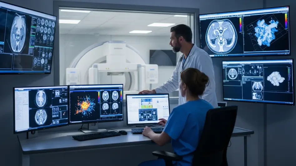
Imaging Precision and AI Integration
Advanced Detection: The NanoPanel Precise System
The technical core of the system revolves around the NanoPanel Precise 4cm XD detector, a component that integrates artificial intelligence directly into the data acquisition phase. This detector is capable of capturing anatomical details as small as 0.25mm, providing a level of clarity that is essential for diagnosing delicate structures such as those found in the inner ear or the complex joints of the musculoskeletal system. By capturing such fine detail, the system allows radiologists to identify tiny fractures or subtle vascular changes that might be missed by less advanced equipment. The AI integration works in real-time to optimize how the detector interprets incoming signals, ensuring that the highest possible resolution is achieved without unnecessarily increasing the radiation dose. This balance of precision and safety is what distinguishes the platform from its predecessors. Radiologists can now rely on a higher level of diagnostic confidence, especially when dealing with difficult pathologies.
Image Purity: Reducing Scatter and Artifacts
To further refine the quality of the images produced, the system utilizes a specialized two-dimensional anti-scatter grid that is designed to minimize visual interference and artifacts. Image scatter is a common problem in CT scanning, particularly when imaging larger patients, as it can blur the edges of internal organs and obscure important diagnostic details. The implementation of this advanced grid technology ensures that the images remain sharp and clear, regardless of the patient’s physical size or the complexity of the scan. This focus on image purity is complemented by the AI-driven noise reduction algorithms that work to smooth out the data while preserving the sharp edges of anatomical structures. The result is a clean, highly legible image that facilitates a more accurate interpretation by the medical staff. By addressing the physical causes of image degradation, the platform provides a more consistent output across all patient demographics. This consistency is vital for maintaining high diagnostic standards.
Specialized AI Suites: Automated Clinical Intelligence
The Rembra platform includes a comprehensive suite of AI-driven tools that are designed to automate the most repetitive and time-consuming tasks faced by imaging technicians and radiologists. These specialized applications, such as the Trauma and Brain suites, can automatically label vertebrae or identify rib fractures within seconds of the scan being completed. In stroke cases, where the phrase “time is brain” is a literal medical reality, these tools can provide an immediate preliminary analysis that highlights areas of concern for the attending physician. By taking over these routine tasks, the AI allows the human experts to spend more of their time on complex decision-making and patient interaction. This automation also reduces the risk of human error that can occur during long shifts or in high-stress emergency situations. The software is designed to be intuitive, integrating seamlessly into the existing digital ecosystem of the hospital to ensure that data flows quickly between clinical departments.
Clinical Automation: Streamlining the Gantry Workflow
Operational efficiency is further improved by the inclusion of patient-side controls that allow the technician to manage the entire setup process directly at the gantry. Traditionally, technicians had to move back and forth between the control room and the patient, which could lead to delays and a loss of focus on the patient’s immediate comfort and safety. With the new interface, the staff can remain at the patient’s side during the critical moments of preparation, ensuring that the alignment and positioning are perfect before the scan begins. This proximity allows for better communication with the patient, which can be especially helpful for those who are anxious or in significant pain. The controls are designed with a simplified layout that prioritizes the most common functions, reducing the amount of training required for new staff members. By streamlining the physical interaction with the machine, the system helps to create a more calm and efficient environment in the imaging suite.
Performance Metrics and Operational Value
Safety Metrics: Dose and Noise Reduction
The efficiency of the system is perhaps most evident in its ability to deliver an 80% reduction in radiation dose while simultaneously decreasing image noise by 85% compared to older hardware. This achievement is a direct result of the iterative reconstruction algorithms and the precision of the new detector technology working in tandem. Low-dose imaging is a critical priority for modern healthcare, as it reduces the long-term risks associated with radiation exposure, which is particularly important for pediatric patients or individuals who require frequent follow-up scans. Despite the lower radiation levels, the images produced are clearer and more detailed than ever before, proving that safety does not have to come at the expense of diagnostic power. This reduction in noise means that radiologists do not have to struggle with grainy images, allowing them to make faster and more definitive calls on the patient’s condition. These improvements translate to better outcomes and efficient patient management.
Operational Speed: High-Speed Data Reconstruction
Speed is another area where the system excels, with the capacity to reconstruct up to 106 images per second and transmit them to the hospital network almost instantly. This rapid processing is enabled by zero-click technology, which automates the transfer and preparation of data so that images are ready for review by the time the patient is moving off the table. In an emergency scenario, every second saved in the imaging process can lead to faster surgical intervention or the quicker administration of life-saving medications. The system effectively removes the waiting period that often occurs while large data sets are being processed by the computer. This immediate availability of information allows the medical team to maintain their momentum during a critical case, moving seamlessly from diagnosis to treatment. The high-speed reconstruction also ensures that the gantry is cleared quickly for the next patient, maximizing the overall throughput. This speed is essential for managing trauma centers.
Long-Term Value: Engineering for Sustainability
From a long-term operational perspective, the Rembra CT was engineered to provide a service life of up to 20 years, making it a sustainable investment for hospitals looking to modernize their infrastructure. The design took into account the need for durability in varied environments, including the ability to function reliably in high-altitude locations where atmospheric pressure can affect sensitive electronics. This longevity is supported by a modular architecture that allows for software updates and hardware upgrades as medical technology continues to evolve. Hospitals that chose to integrate this platform found that it could transition seamlessly between the high-volume demands of diagnostic radiology and the extreme precision required for radiation therapy planning. This versatility allowed healthcare administrators to consolidate their imaging needs into a more efficient footprint, reducing the need for multiple specialized machines. The focus on long-term reliability ensured that the technology remained a stable asset.
Practical Implementation: Insights and Next Steps
Healthcare administrators who prioritized these advanced imaging capabilities realized significant improvements in both patient throughput and clinical accuracy across their emergency departments. The integration of AI-driven automation effectively reduced the workload on their technical staff, allowing for a more focused approach to patient care during high-stress shifts. Organizations that implemented the platform were encouraged to develop specific protocols that maximized zero-click processing features to further decrease wait times. These early adopters observed that the combination of low-dose imaging and high-resolution output set a new benchmark for safety and diagnostic excellence. Moving forward, the industry was advised to consider how such versatile hardware could be leveraged across multiple departments to optimize expenditure. The success of the rollout demonstrated that investing in intelligent, durable hardware was an effective strategy for meeting the demands of modern care. Future developments were anticipated to build upon this foundation.