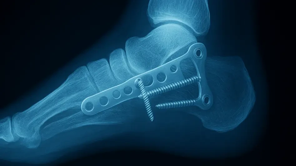

Surgeons often struggle with the dense overlay of metal plates and screws on postoperative images, which frequently hide subtle but critical hardware complications. This diagnostic hurdle is particularly evident in cases involving the calcaneus, an irregularly shaped bone responsible for supporting the total weight of the human body. When this heel bone undergoes a comminuted fracture—typically due to severe high-energy trauma like falls from significant heights—the surgical reconstruction involves complex fixation methods. Radiologists face an uphill battle when interpreting subsequent X-rays, as the two-dimensional nature of the imaging compresses a three-dimensional labyrinth of metal hardware and healing tissue. These images are often so cluttered that distinguishing between a stable recovery and a misplaced screw penetrating a joint space becomes a matter of intense clinical debate. Consequently, clinicians frequently rely on expensive CT scans to achieve the necessary clarity, which increases costs and radiation exposure for patients.
Rigorous Methodology: Multi-Reader Study Design
To address these persistent clinical bottlenecks, researchers Shaobin Wang and Hai Li spearheaded a multicenter, multi-reader study designed to evaluate the efficacy of multimodal large language models in musculoskeletal radiology. The investigation utilized GPT-5 as a diagnostic co-pilot, aiming to determine if AI could provide the necessary support to increase both the speed and accuracy of X-ray interpretations. The study involved six radiologists with varying levels of professional experience, ensuring that the results would be applicable to a wide range of hospital settings and expertise levels. These participants analyzed 73 specific cases of postoperative calcaneal fixation, resulting in a dataset of 438 individual observations. To establish a definitive ground truth, every X-ray was meticulously compared against a pragmatic reference standard, which consisted of a postoperative CT scan interpreted by a consensus of experts. This high standard allowed the team to measure AI performance against the most reliable clinical data currently available.
The experimental framework was structured into two distinct phases, separated by a washout period of four weeks to ensure that the radiologists did not retain specific memories of the cases they had previously reviewed. During the initial session, the doctors worked entirely unaided, following their standard clinical protocols to record findings and confidence levels. This served as a baseline for comparison against the second phase, where GPT-5 was introduced into the workflow. In the AI-assisted session, the readers were provided with pre-generated draft reports and structured checklists created by the model. GPT-5 analyzed both the pixel data of the images and relevant textual clinical context to highlight potential hardware issues or healing anomalies. The radiologists were then tasked with reviewing, editing, and finalizing these suggestions. This collaborative approach aimed to combine the nuanced judgment of a human clinician with the massive data-processing capabilities of a modern large language model.
Diagnostic Performance: Enhancing Accuracy and Sensitivity
The quantitative results of the study provided a compelling argument for the integration of GPT-5 into routine orthopedic imaging workflows. When assessing cases that required clinical escalation—those where medium- to high-risk complications were present—the diagnostic accuracy of the participating radiologists saw a significant improvement. In the unaided sessions, the accuracy rate hovered at 71.5 percent, reflecting the inherent difficulty of interpreting these complex images. However, when the radiologists utilized the AI-generated checklists, this figure rose to 80.1 percent. This statistical jump represents a near-doubling of the odds that a radiologist’s final decision would align perfectly with the expert CT consensus. By bridging the gap between traditional 2D X-rays and the granular detail typically reserved for 3D imaging, the AI model essentially provided a higher level of insight without the need for additional radiation exposure. This suggests that the model can help standardize the quality of care across different medical facilities.
A particularly vital metric identified during the research was the dramatic increase in diagnostic sensitivity, which is the ability of a test to correctly identify a problem when it is truly present. The study found that sensitivity increased from 54.2 percent to 79.2 percent when AI assistance was employed. This finding indicates that the GPT-5 draft reports functioned as an essential safety net, drawing the radiologist’s attention to subtle hardware misplacements or fracture lines that might otherwise be overlooked in a cluttered visual field. While there was a minor trade-off in specificity—the ability to correctly identify a stable recovery—which dropped from 85.0 percent to 80.9 percent, the clinical implications are largely positive. In the context of heel fractures, missing a major complication can lead to permanent disability and chronic pain, making a slight increase in false alarms a manageable risk compared to the danger of false negatives. The model proved most effective at flagging general complications, though high-risk cases still required a CT scan.
Clinical Impact: Workflow Efficiency and Confidence Calibration
Beyond the improvements in diagnostic precision, the study highlighted a transformative shift in the efficiency of the radiological workflow. The mean reading time per case plummeted from 378.6 seconds when the doctors worked alone to just 98.4 seconds with the assistance of GPT-5. In a modern, high-pressure hospital environment, completing a complex postoperative report in roughly a quarter of the usual time could have profound implications for patient throughput and staff well-being. By automating the preliminary drafting of reports and the organization of findings into a structured format, the AI allows clinicians to bypass the more tedious aspects of documentation. This efficiency gain enables radiologists to focus their cognitive energy on high-level clinical decision-making and patient interaction rather than the manual labor of report generation. For trauma centers dealing with high volumes of orthopedic cases, this technology could significantly alleviate the administrative burden that frequently contributes to burnout among medical personnel.
Another critical aspect of the study focused on confidence calibration, which was measured using the Brier score to assess how well a doctor’s self-reported confidence aligned with their actual accuracy. The researchers observed that the AI-assisted sessions resulted in improved Brier scores, dropping from 0.246 to 0.177. This improvement suggests that the presence of the AI model helped the radiologists become more self-aware regarding the limitations of their own diagnostic conclusions. Rather than just providing answers, the GPT-5 system acted as a cognitive mirror, helping the clinicians feel more certain when they were correct and more appropriately cautious when the visual data was ambiguous. This refinement of human judgment is a key component of patient safety, as it reduces the likelihood of overconfident errors. When clinicians are better calibrated, they are more likely to seek secondary opinions or additional imaging only when truly necessary, leading to a more streamlined and reliable healthcare experience for every patient.
Future Path: Methodological Integrity and Considerations
The research team received substantial praise for their methodological integrity, specifically for adhering to transparency standards such as the STARD-AI and the CLAIM checklist. By making their analysis code available and utilizing complex statistical models to account for reader variability, they have established a rigorous blueprint for future medical AI evaluations. However, the authors also remained cautious, acknowledging certain limitations inherent in the study design, such as the potential for an order effect. Since the AI-assisted sessions always followed the unaided sessions, there is a possibility that some performance improvements were the result of the radiologists becoming more familiar with the specific case files. Although the four-week washout period and randomized case order were intended to mitigate this bias, the researchers noted that the study was not fully counterbalanced. This realization led the authors to conclude that while the findings are exceptionally promising, they should be treated as a compelling candidate for clinical adoption.
Looking forward, the medical community established a clear pathway for integrating these multimodal cognitive partners into daily practice to ensure higher standards of orthopedic care. The study demonstrated that the most effective use of AI was not as a replacement for human expertise but as an interactive assistant that supported existing clinical workflows. Hospitals and clinics were encouraged to begin exploring pilot programs that utilize GPT-5 for preliminary report drafting and structural checklists, particularly in high-stakes trauma scenarios. Further prospective and randomized trials across international institutions were initiated to confirm these initial findings and to refine the interaction between radiologists and AI models. This evolution in diagnostic technology pointed toward a future where the management of complex injuries, like calcaneal fractures, became significantly more predictable and efficient. By focusing on the synergy between human judgment and automated data analysis, healthcare providers successfully reduced the margin for error.