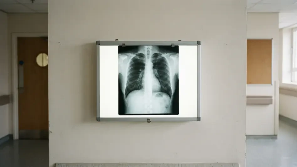

The interpretation of chest X-rays remains inherently difficult because medical lesions, such as localized nodules or pleural effusions, vary drastically in scale and appearance within a single image. Modern medicine is increasingly turning to computer-aided diagnosis to manage the overwhelming volume of medical imaging required for patient care. Researchers have recently introduced PMSL-YOLO, an innovative framework designed to detect multiple thoracic diseases within chest radiographs using a lightweight architecture. By building upon the YOLOv8n model, this study seeks to provide high-tier accuracy without requiring expensive, high-end hardware that often limits AI in smaller clinics or resource-constrained regions. This advancement is particularly timely as healthcare systems look for ways to integrate AI without incurring massive infrastructure costs. Efficiency ensures that sophisticated support is not reserved for elite hospitals, allowing for wider implementation across various diagnostic settings.
Bridging the Gap in Clinical Hardware Accessibility
The Necessity for Efficient Diagnostic Tools
High-performance deep-learning models typically demand substantial computational resources, creating a technological barrier for many healthcare facilities. While powerful AI exists, its reliance on costly graphics processing units makes it inaccessible to community clinics with limited budgets. The development of PMSL-YOLO addresses this disparity by optimizing a “nano” architecture to perform at levels previously reserved for much heavier models, potentially democratizing advanced diagnostic assistance. In many rural or underfunded settings, the inability to purchase high-end servers has meant that patients do not benefit from the latest breakthroughs in imaging technology. By focusing on a lightweight framework, researchers ensure that the computational overhead is low enough to run on standard hardware. This shift represents a move toward inclusive technology that prioritizes clinical utility over model size, facilitating a more equitable distribution of global resources.
Enhancing Radiologist Productivity and Safety
Furthermore, the heavy workload faced by radiologists in contemporary practice leads to significant fatigue, which increases the likelihood of human error during image interpretation. Chest X-rays are the most common tool for screening thoracic abnormalities, yet identifying multiple coexisting diseases remains a complex task for even the most experienced clinicians. Automated systems like PMSL-YOLO provide a critical safety net, functioning as a second set of eyes that does not tire or lose focus during a long shift. For residents and early-career physicians, this support is even more vital, as it helps bridge the gap in clinical experience by highlighting subtle markers that might otherwise be overlooked. By integrating these efficient diagnostic tools into the daily workflow, hospitals can mitigate the risks associated with clinician burnout while improving the speed of diagnosis. This dual benefit of reducing human strain is essential for maintaining high standards of care across all regions.
Advancing Detection Through Multi-Scale Training
Innovation in Resolution and Data Processing
The core innovation of this framework is Progressive Multi-Scale Learning, which moves away from the traditional fixed-resolution training method. By starting with lower resolutions to teach the model broad anatomical structures and incrementally increasing to higher resolutions, the system learns to “zoom in” on tiny nodules without losing the context of the entire chest cavity. This guided evolution allows the lightweight model to refine its feature extraction capabilities, significantly improving the detection of varied lesion sizes without increasing complexity. In standard models, small abnormalities often vanish during the downsampling process, but the PMSL approach ensures that these features are preserved and prioritized as the resolution scales up. This methodology mirrors the way a human radiologist might scan an image for general symmetry before focusing on specific regions. By mimicking this progression, the AI achieves a nuanced understanding of thoracic pathology.
Specialized Augmentation for Clinical Robustness
To further bolster performance, the researchers utilized specialized data augmentation techniques such as image blending and mosaic augmentation to simulate various clinical scenarios. These methods help the AI recognize objects in different contexts and adapt to varying image qualities produced by different X-ray machines. By adjusting brightness, contrast, and geometric positioning, the model becomes robust against the real-world inconsistencies found in hospital imaging departments, particularly when identifying subtle, localized lesions. This is crucial because real-world X-rays are rarely as clean as laboratory datasets; variations in patient positioning or equipment calibration can often confuse less robust algorithms. The inclusion of these augmentation strategies ensures that the PMSL-YOLO framework remains reliable across different hardware brands and technical setups. Such adaptability is a prerequisite for any tool intended for widespread clinical deployment.
Empirical Results and Real-World Application
Rigorous testing on benchmark datasets revealed that this progressive approach offers a significant statistical advantage, achieving a recall rate of over 92%. In a medical context, high recall is vital because it ensures that fewer actual diseases are missed, reducing the risk of life-threatening false negatives. The results indicate that the strategy not only makes the AI more accurate but also makes it a more reliable safety net for radiologists working in fast-paced environments. Precision also saw a marked improvement, rising to over 94%, which demonstrates that the model identifies genuine abnormalities with high confidence. These metrics suggest that the PMSL-YOLO framework can effectively handle the complexities of multi-disease detection where several different pathologies may overlap in a single image. Achieving such results on a lightweight architecture is a major milestone, proving that efficiency does not have to come at the expense of reliability or safety.
Actionable Steps for Future Clinical Integration
The development of the PMSL-YOLO framework demonstrated that lightweight neural networks can successfully navigate the complexities of multi-disease detection in chest radiographs. By prioritizing progressive multi-scale learning, the researchers proved that high-tier diagnostic accuracy was achievable without the need for prohibitive hardware investments. This study validated the potential for AI to serve as an accessible safety net in diverse clinical settings, effectively bridging the gap between resource-rich and resource-constrained environments. Healthcare administrators who reviewed these findings noted that efficient architectures offered a sustainable path toward AI integration. Clinicians who participated in pilot programs provided feedback on real-world utility, ensuring that subsequent refinements addressed actual workflow bottlenecks. By adopting these scalable solutions, the medical community established a firm foundation for more equitable healthcare delivery for patients.