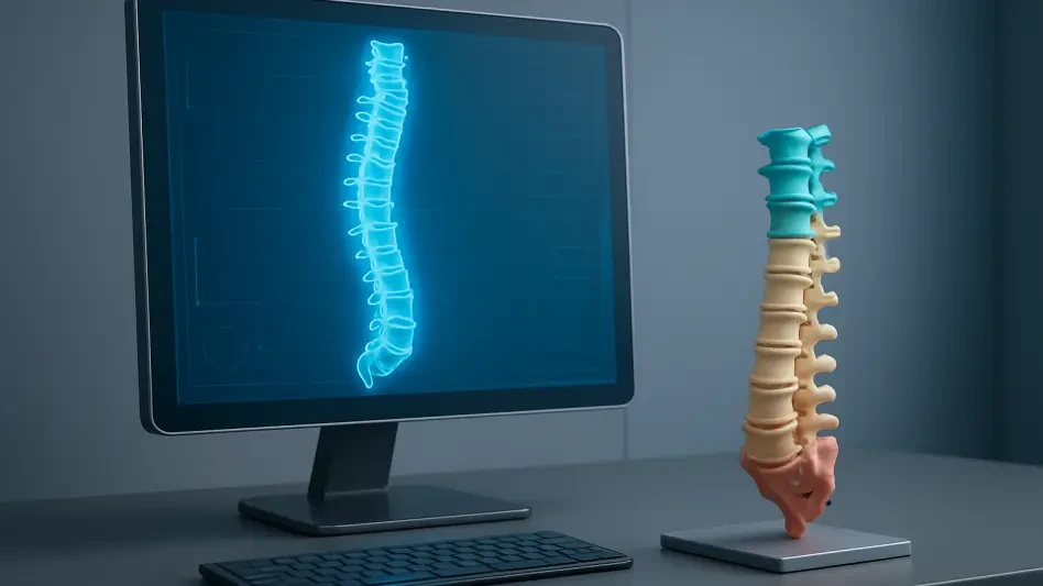

A surgeon standing in a modern operating room can now physically rotate a patient’s translucent ribcage or manipulate a life-sized model of a failing heart before ever picking up a scalpel. This profound shift from viewing flat images on a lightbox to interacting with high-fidelity physical replicas marks a departure from a century of traditional medical practice. In this landscape, the anatomical digital twin has moved from the laboratory to the sterile field, effectively closing the gap between diagnostic theory and the tactile reality of the human body. As procedures become increasingly complex, the ability to rehearse an operation on a replica that mirrors every nuance of a patient’s unique internal landscape has become a standard of excellence in the most advanced medical centers.
In 2026, the transition toward a more personalized surgical experience has reached a critical tipping point. High-resolution imaging combined with rapid additive manufacturing allows clinical teams to move beyond the limitations of generic anatomical knowledge. This revolution is not merely about the aesthetics of a 3D model; it is about the tangible reduction of risk, the shortening of recovery times, and the elimination of the guesswork that has historically plagued the most intricate surgeries. By transforming data into physical form, medicine has entered an era where every patient receives a treatment plan as unique as their own DNA.
The End of Surgical Guesswork in a Three-Dimensional Era
The human body does not exist in two dimensions, yet for decades, surgeons have been forced to translate flat CT scans and MRIs into a mental 3D map while holding a scalpel. This cognitive leap is rapidly becoming a relic of the past as high-fidelity anatomical reconstructions move from the screen to the sterile field. Today, a surgeon can hold a patient’s heart in their hands before the first incision is even made, transforming “unconventional anatomy” from a surgical surprise into a premeditated plan.
The elimination of spatial ambiguity allows for a level of rehearsal that was previously impossible. When a surgeon interacts with a physical replica of a tumor intertwined with major arteries, they can identify the safest entry points and angles of approach without the pressure of an active clock. This premeditated approach effectively lowers the cognitive burden on the surgical team, shifting their focus from spatial interpretation to technical execution. Consequently, the operating room becomes an environment of controlled action rather than one of discovery.
Bridging the Gap Between Imaging and the Operating Table
Medical imaging has long provided the data necessary for diagnosis, but traditional methods often fail to capture the spatial complexity of intertwined blood vessels, airways, and neurological pathways. This gap creates a reliance on intraoperative decision-making, which can increase the time a patient spends under anesthesia. As healthcare trends shift toward personalized medicine, the demand for patient-specific solutions has turned 3D modeling from an experimental novelty into an essential component of the modern surgical workflow.
Bridging this digital-to-physical divide requires more than just high-resolution data; it requires a seamless integration of radiology and surgery. Modern workflows ensure that the digital insights gained during the scanning phase are directly applicable to the physical challenges of the surgery. By using these models as a bridge, medical teams can anticipate structural anomalies that might otherwise only become visible once the patient is on the table. This foresight is particularly critical in pediatric cases, where small-scale anatomy leaves very little room for error.
The Technical Evolution of Anatomical Reconstruction
The transition from raw data to a physical or virtual model is a rigorous process known as segmentation, where flat images are layered to create a voluminous digital twin. Specialists now use advanced software to isolate specific structures, such as brain ventricles or intricate bone fragments, transforming them into digital renderings within minutes. This speed is vital for trauma cases where time is the most precious resource.
Furthermore, surgeons are no longer limited to a single view; they can choose between sterilized 3D prints for tactile feedback, VR environments for immersive exploration, or “video-game-style” interactive interfaces. Modern hardware allows for the production of physical replicas within a single day, ensuring that even urgent cases can benefit from bespoke surgical planning. This diversification of media means that whether a surgeon needs to feel the texture of a bone or fly through a virtual artery, the technology adapts to their specific sensory requirements.
Expert Perspectives on Institutional Integration
While individual “physician-enthusiasts” initially pioneered these tools, the industry is moving toward a centralized, in-house service model to ensure scalability and precision. Leading hospitals are establishing dedicated 3D printing departments that serve pediatric cardiology, neurosurgery, and orthopedics under one roof. This centralized hub approach allows for the cross-pollination of ideas and the standardization of quality across multiple specialties.
Experts emphasize that the best models are born from a partnership between imaging specialists and surgeons, allowing for customized depth markers and color-coded structures. Beyond the operating room, physicians use these physical models to bridge language and education gaps, helping families visualize complex procedures with a level of clarity that a computer screen cannot provide. When a parent can see and touch a model of their child’s heart, the abstract fear of surgery is replaced by a concrete understanding of the solution.
Strategies for Optimizing Surgical Efficiency and Safety
Implementing 3D technology is not just about the hardware; it is about applying specific frameworks to improve patient outcomes and hospital economics. Surgeons can use 3D prints to pre-bend plates and manipulate surgical guides before entering the OR, eliminating the need for on-the-fly adjustments. By resolving technical challenges in a virtual or tactile rehearsal, teams can shorten complex operations by 30 to 90 minutes, directly reducing the window for potential complications.
Utilizing bespoke templates with built-in depth markers ensures millimeter precision, which lowers the overall cost per procedure by reducing the likelihood of revision surgeries. These strategies represent a shift toward industrial-level precision within the artisanal field of surgery. By front-loading the technical problem-solving, hospitals can maximize the throughput of their operating rooms while simultaneously elevating the safety profile for every individual patient.
The successful implementation of these 3D protocols rested on the development of rigorous quality control measures that governed the translation of digital data into surgical reality. Hospitals prioritized the creation of multidisciplinary committees that included engineers, radiologists, and clinicians to oversee the accuracy of every printed guide. This collaborative structure ensured that technical innovation remained grounded in clinical necessity, preventing the technology from becoming a mere executive ornament. Moving forward, the industry addressed the need for standardized reimbursement models, allowing smaller community hospitals to adopt these life-saving tools without incurring prohibitive costs. By treating the patient’s anatomy as a navigable map rather than a mystery, the surgical community definitively shifted the focus from improvisation to total precision.