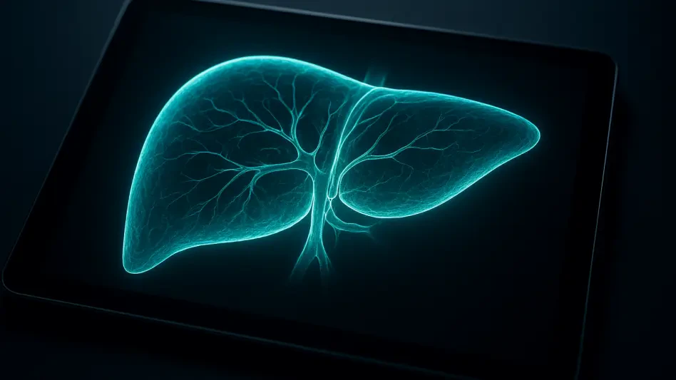

Standard deep learning models often produce anatomically impossible shapes, such as disconnected vascular pathways, which are technically correct by pixel counts but clinically useless for actual surgical planning. The current landscape of medical imaging suffers from a distinct imbalance where high-resolution CT scans are plentiful, yet the expert-level labels required to train artificial intelligence remain prohibitively expensive and time-consuming to produce. Radiologists frequently spend hours manually delineating the borders of the liver, spleen, and intricate vascular networks, creating a bottleneck that limits the rapid deployment of automated diagnostic tools. In response to this challenge, a research team from the Fourth Clinical College of Henan Medical University has introduced URDT-Net. This semi-supervised framework is designed to bridge the data gap by utilizing massive pools of unlabeled images, effectively learning from the vast majority of clinical data that previously went unused in training.
Dual-Teacher Systems: Robust Pseudo-Label Generation
The architectural foundation of URDT-Net rests on a sophisticated dual-teacher paradigm that moves beyond the limitations of standard student-teacher models. Most existing systems utilize a single teacher network to generate pseudo-labels for unlabeled data, but this often results in a narrow perspective that fails to capture the complexity of abdominal anatomy. By employing two distinct exponential-moving-average teachers, URDT-Net creates a collaborative learning environment where different aspects of an image are prioritized simultaneously. One teacher focuses on the overall volumetric shape and coarse morphology of the organs, while the complementary teacher is tuned to recognize high-frequency details such as sharp boundaries and fine edges. This diversity of viewpoints allows the system to produce a fused pseudo-label that is far more accurate and comprehensive than what any individual model could achieve alone, ensuring the student network receives a high-quality training signal.
To further refine the quality of these automated labels, the researchers implemented an uncertainty-ranked retention mechanism that acts as a rigorous gatekeeper for incoming data. Semi-supervised learning is notoriously vulnerable to noise propagation, where a model accepts a wrong guess as truth and subsequently reinforces that error throughout the training cycle. URDT-Net addresses this by utilizing Monte Carlo sampling to assess the confidence level of its own predictions in real-time. By ranking pseudo-labels based on their statistical reliability, the framework filters out high-uncertainty guesses and only permits the most certain predictions to influence the model’s parameters. This selective learning process is particularly crucial for smaller or low-contrast structures like the pancreas or adrenal glands, where the margin for error is razor-thin. Consequently, the system avoids the pitfalls of misinformation, turning the abundance of unlabeled clinical data into a reliable asset rather than a source of potentially dangerous algorithmic confusion.
Topological Consistency: The Preservation of Anatomical Truth
A primary innovation within the URDT-Net framework is its strict adherence to topological consistency, which ensures that the segmented outputs respect the physical laws of human biology. Traditional voxel-wise loss functions often fail to penalize results that are mathematically close but biologically nonsensical, such as a solid liver appearing with internal gaps or a hepatic vein manifesting as a series of isolated spheres. To rectify this, the researchers integrated an organ-aware topology objective that utilizes specialized constraints to maintain structural integrity. One such constraint, the soft morphological-survival term, specifically prevents the formation of unrealistic internal voids within solid organs by encouraging the model to maintain a continuous volumetric mass. This approach shifts the priority from simple pixel matching to a more holistic understanding of how organs occupy space, ensuring that the final segmentation is not just a collection of classified points but a functionally coherent representation of the patient’s internal anatomy.
The preservation of connectivity is equally vital for tubular structures, which are handled through a dedicated centerline Dice objective. This mechanism prioritizes the extraction of the skeletonized path of blood vessels, ensuring that the model maintains the functional continuity of the vascular tree regardless of the vessel’s diameter or contrast level. Furthermore, the system incorporates an inter-class ambiguity term that specifically manages the high-stakes boundaries between adjacent organs in the crowded abdominal cavity. By explicitly modeling the transition zones where the liver might meet the kidney or the stomach, URDT-Net significantly reduces the likelihood of segmentation bleeding, where the pixels of one organ are incorrectly attributed to its neighbor. This level of topological precision is essential for surgical navigation and radiation therapy planning, where even a small overlap or a disconnected pathway could lead to errors in clinical judgment or treatment delivery during high-pressure medical procedures.
Efficiency Metrics: Budget-Aware Early-Exit Mechanisms
While accuracy is paramount, the practical application of 3D deep learning models in busy hospital environments is often hindered by significant computational demands and latency issues. URDT-Net addresses these real-world constraints through an innovative budget-aware design featuring an intermediate segmentation head that facilitates early-exit capabilities. During the inference process, the model evaluates its own confidence at a halfway point within the neural network layers. If the intermediate prediction meets a predefined threshold of certainty, the system terminates the calculation early and provides the result immediately, bypassing the remaining layers of the architecture. This mechanism allows the framework to process easy cases with minimal resource expenditure while reserving its full computational power for the more complex or ambiguous scans that require deeper analysis. This dynamic allocation of resources ensures that the system remains highly responsive, providing radiologists with rapid results without sacrificing the depth of focus required for difficult diagnoses.
The performance of URDT-Net was validated against the rigorous FLARE22 benchmark, demonstrating clear superiority over contemporary semi-supervised methods. In comparative trials, the framework achieved a mean Dice similarity coefficient of 0.8543, surpassing competitors that relied on simpler data augmentation or single-teacher architectures. Perhaps more importantly, the system showed a marked reduction in the 95th-percentile Hausdorff distance, falling from 15.62 mm in standard models to 13.42 mm, which indicates far fewer large-scale errors at organ boundaries. Efficiency metrics were equally impressive, as the early-exit mechanism allowed the system to conclude processing on nearly 39 percent of the test volumes well before reaching the final layer. This led to a 25 percent reduction in total latency, dropping the average processing time per volume from over six seconds to just 4.6 seconds. These gains in both precision and speed suggest that the framework is exceptionally well-suited for the high-throughput demands of modern radiology departments and emergency rooms.
Implementation Success: Perspectives on Clinical Integration
The successful development and validation of URDT-Net highlighted a significant shift toward more practical and anatomically aware artificial intelligence in the medical field. By moving beyond simple data-labeling paradigms and embracing a multi-teacher approach that prioritized structural continuity, the research team provided a blueprint for more reliable diagnostic assistants. The study demonstrated that the initial investment in a more complex training phase was justified by the resulting gains in inference speed and topological accuracy at the clinical bedside. Clinicians who utilized these tools observed that the reduction in manual correction time for vascular pathways and organ boundaries significantly streamlined their daily workflows. Furthermore, the ability to leverage massive amounts of unlabeled data from 2026 onwards suggested that AI performance would continue to improve as more digital records became available, provided that the underlying models possessed the necessary common sense regarding human anatomy and the computational efficiency to be useful.
Looking ahead, the principles established by the URDT-Net framework offered a clear path for expanding semi-supervised learning into other complex imaging domains, such as cardiac or neurosurgical planning. Healthcare organizations that prioritized the infrastructure necessary to handle high-volume unlabeled data streams found that budget-aware mechanisms were essential to their specific hardware limitations. The transition toward systems that understood the connectivity of the human body, rather than just its pixel intensity, marked a turning point in the trust levels between radiologists and automated software. As these tools became more integrated into standard practice, the emphasis shifted toward developing adaptive systems that learned from localized clinical feedback, further bridging the gap between generalized algorithmic performance and the unique needs of individual patients and specialized surgical teams. The team recommended that future iterations explore multi-modal data integration to enhance the precision of these structural maps even further.