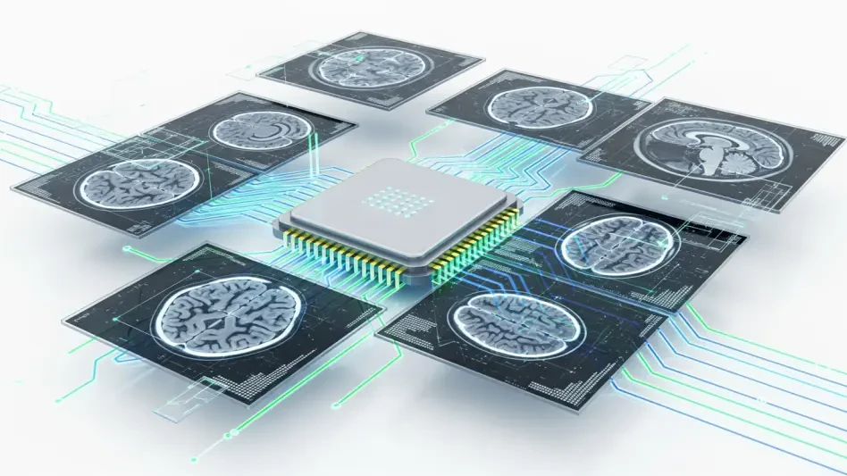

The persistent challenge of accurately diagnosing Parkinson’s disease in its earliest stages has long stymied neurologists who must often rely on subjective motor assessments or prohibitively expensive imaging techniques like magnetic resonance imaging. While these standard diagnostic pathways provide some clarity, they frequently miss subtle neurological changes that occur years before the onset of tremors or significant bradykinesia. This diagnostic gap has fueled a surge in research focused on more accessible, objective methodologies, leading to the development of a sophisticated computational framework that leverages the power of artificial intelligence. By integrating machine learning with ultrasound technology, scientists have created a path toward rapid, cost-effective screening that could fundamentally alter the landscape of neurodegenerative care. This breakthrough shifts the focus from waiting for visible symptoms to identifying microscopic shifts in brain structure, ensuring that interventions can begin much earlier in the disease cycle than previously possible.
Sonography and Artificial Intelligence: Overcoming Technical Hurdles
Transcranial Sonography (TCS) has emerged as a promising alternative to traditional neuroimaging due to its non-invasive nature and relative ease of use in clinical settings. Unlike the bulky and expensive MRI machines found only in major medical centers, TCS devices can be used at the bedside to visualize the substantia nigra, a brain region central to Parkinson’s pathology. However, the practical utility of TCS has historically been limited by significant visual noise and artifacts caused by the varying thickness of the human skull, which often obscures the very structural changes clinicians need to see. This inconsistency has led to high variability in interpretation, where even experienced sonographers might struggle to differentiate between healthy aging and early disease markers. Consequently, the reliance on human eye interpretation alone has prevented TCS from becoming a primary diagnostic standard, despite its potential for widespread screening in various healthcare environments.
To address these inherent limitations, researchers have introduced a specialized artificial intelligence framework designed to process and clarify these complex ultrasound images with unprecedented precision. This system employs advanced signal processing techniques to filter out the interference caused by bone density and soft tissue, allowing the underlying neurological patterns to emerge with greater clarity. By training on vast datasets of validated brain scans, the AI can detect minute echogenicity changes in the midbrain that are virtually invisible to human observers. This capability transforms a traditionally subjective imaging tool into a highly objective diagnostic instrument, capable of providing consistent results regardless of the operator’s experience level or the specific hardware used. The integration of such robust computational power ensures that the diagnostic process is not only faster but also significantly more reliable, laying the groundwork for a new standard in routine neurological assessments.
Sophisticated System Design: The Stacked Multi-Classifier Approach
The mechanical heart of this diagnostic advancement lies in its unique stacked multi-classifier framework, which represents a significant leap over conventional single-algorithm AI models. In this architecture, multiple base models operate in parallel, each specialized in analyzing different aspects of the ultrasound data, such as texture, intensity, or geometric symmetry within the brain regions. These individual outputs are then synthesized by a high-level meta-classifier that weighs the evidence from each source to arrive at a final diagnostic conclusion. This tiered learning approach is particularly effective for handling the inherent complexity of biological data, as it allows the system to cross-reference multiple features simultaneously and reduce the likelihood of false positives. By distributing the computational workload across specialized sub-units, the framework achieves a level of accuracy that matches or exceeds the performance of traditional radiologists in most cases.
Beyond its purely visual analysis, the framework excels through its use of multi-modal data fusion, a process that integrates diverse streams of information to create a holistic patient profile. Rather than relying solely on ultrasound images, the AI incorporates clinical variables such as the patient’s age, specific motor symptoms, and comprehensive medical history into its decision-making matrix. This holistic view is crucial because Parkinson’s disease manifests differently across various demographics, and symptoms can often overlap with other neurological conditions. By combining structural imaging with functional clinical data, the model develops a multidimensional diagnostic signature that provides a far more complete picture of the patient’s health than any single test could offer. This synthesis of information ensures that the final diagnosis is grounded in both the biological reality of the brain’s structure and the practical reality of the patient’s clinical physical experience.
Clinical Transparency: Bridging the Gap Between AI and Medicine
A significant barrier to the adoption of artificial intelligence in healthcare has been the black box problem, where sophisticated algorithms provide answers without explaining the underlying reasoning. To overcome this hurdle, the new framework incorporates explainability features that allow clinicians to see exactly which features of an image or which specific symptoms led to a particular diagnosis. Through the use of saliency maps and heatmaps, the system highlights the areas within the substantia nigra or other brain structures that exhibit the most significant abnormalities. This visual evidence provides a bridge between the computer’s logic and the doctor’s clinical expertise, allowing for a collaborative diagnostic process rather than a blind reliance on automated output. When a physician can verify the AI findings against their own observations, it builds the necessary trust required for these tools to move into everyday hospital workflows.
This focus on interpretability also plays a vital role in medical education and the refinement of diagnostic criteria themselves. As the AI identifies novel patterns or subtle markers that were previously unrecognized by the medical community, researchers can analyze these insights to gain a deeper understanding of Parkinson’s pathology. The framework essentially acts as a powerful microscope that not only sees more clearly but also points out what is most important to look at, thereby accelerating the discovery of new biomarkers. Furthermore, providing a clear rationale for every diagnosis is essential for meeting the rigorous regulatory and ethical standards required in modern medicine. By prioritizing transparency, the developers of this framework have ensured that the technology is not only technically superior but also socially and ethically responsible, ensuring that the tool is both useful to the provider and safe for the patient.
Global Accessibility: Expanding the Reach of Neurological Expertise
One of the most compelling advantages of this AI-driven ultrasound approach is its potential to democratize high-quality neurological care across the globe. Traditional neuroimaging technologies like MRI and PET scans are often concentrated in wealthy urban areas, leaving millions of people in rural or underserved regions without access to early diagnostic services. In contrast, ultrasound machines are portable, relatively inexpensive, and require significantly less infrastructure to operate, making them ideal for deployment in community clinics. By imbuing these affordable devices with high-level diagnostic intelligence, the framework effectively exports the expertise of world-class neurologists to any location where a basic ultrasound probe is available. This shift has the potential to drastically reduce the global burden of undiagnosed Parkinson’s disease, ensuring that geography no longer dictates the quality of care.
To ensure that the system is ready for such widespread application, the research team employed rigorous cross-validation techniques and tested the model against diverse datasets from various global populations. This process was designed to prevent overfitting, a common issue where an AI performs exceptionally well on the data it was trained on but fails to generalize to new patients with different physical characteristics. By validating the framework across different types of ultrasound equipment and varying degrees of image quality, the team demonstrated that the system is robust enough to handle the realities of different clinical environments. This reliability is essential for a tool intended for global use, as it must maintain its accuracy regardless of the local technical constraints or the specific demographic being screened. The result is a truly versatile diagnostic tool that bridges the gap between research and practical medical needs.
Evolutionary Diagnostics: Longitudinal Tracking and Broadened Scope
The development team successfully integrated longitudinal data tracking, which allowed the framework to monitor disease progression over extended periods rather than providing a single snapshot in time. By comparing scans and clinical data collected at different intervals, the AI began to identify specific trajectories of decline, offering doctors the ability to predict how a patient condition might evolve in the coming years. This shift from static diagnosis to dynamic monitoring was essential for tailoring personalized treatment plans that were adjusted as the disease moved through different phases. Such a capability proved invaluable in clinical trials, where researchers needed to measure the efficacy of new neuroprotective therapies with high precision. The system provided an objective way to see if a particular intervention was slowing the rate of structural changes in the brain, thereby improving the overall patient outcome.
The successful implementation of this framework also paved the way for its adaptation to other neurodegenerative conditions, such as Alzheimer’s disease and multiple sclerosis. Because the underlying architecture was designed to be modular and scalable, researchers were able to retrain the models on different types of imaging data and clinical markers relevant to these other disorders. This expanded scope transformed the tool from a specialized Parkinson’s diagnostic into a comprehensive platform for neurological health, capable of screening for multiple conditions simultaneously. Furthermore, the commitment to diversifying training datasets ensured that the AI remained equitable and effective for patients of all ethnic backgrounds. As these systems became more integrated into the standard of care, they fostered a new era of personalized medicine where early detection and continuous monitoring became the standard for treating complex diseases.