
The historical reliance on invasive biopsies and fragmented imaging protocols has finally given way to a sophisticated era of medical technology where artificial intelligence and ultrasound merge into a single diagnostic powerhouse. For decades, clinicians struggled with the ambiguity of grainy images, often requiring patients to undergo multiple sessions across different departments to obtain a complete picture of liver health. This inefficient cycle not only delayed critical treatment but also increased the economic burden on healthcare systems worldwide. By 2026, the landscape has shifted toward a data-driven ecosystem where high-resolution anatomical imaging is no longer a standalone tool but part of a comprehensive quantitative analysis. This transformation is driven by the integration of machine learning algorithms that process echoes in real-time, providing immediate clarity on tissue composition. Consequently, the industry is moving away from incremental hardware updates in favor of unified platforms that prioritize clinical accuracy and operational efficiency for every patient.
Bridging the Gap Between Imaging and Diagnostics
Modern diagnostic platforms have effectively collapsed the traditional, multi-step clinical pathway into a single, streamlined encounter that allows for the simultaneous assessment of the liver, spleen, and gallbladder. In the past, a patient might have been scheduled for a standard ultrasound on one day and a specialized elastography session weeks later, leading to fragmented data and prolonged anxiety. Today, advanced software suites allow medical professionals to visualize organ structure while capturing functional data in one continuous workflow. This holistic perspective is particularly vital for managing systemic digestive diseases, as pathologies in the liver frequently correlate with issues in the surrounding vasculature or the spleen. By seeing the entire physiological context at once, hepatologists can make faster, more informed decisions without the need for redundant follow-up appointments. This synchronization of data points ensures that the diagnostic narrative is complete and cohesive from the very first screening.
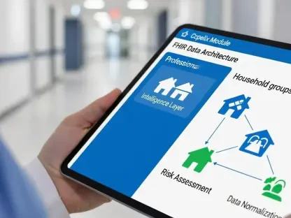
Beyond the walls of centralized imaging departments, the emergence of portable, tablet-based ultrasound units is bringing hospital-grade diagnostics directly to the point of care in community clinics. These compact systems maintain the same AI-enhanced processing power as their larger counterparts, ensuring that the quality of the diagnostic output is not compromised by the physical size of the device. This newfound flexibility allows high-level liver assessments to occur at the bedside or in remote rural areas where access to specialized hepatology centers was previously limited. As healthcare networks strive to meet the diverse logistical demands of a growing population, the ability to deploy sophisticated tools in varied environments becomes a critical factor in public health. Providers can now implement proactive screening programs that catch chronic conditions in their infancy, long before they escalate into emergencies requiring intensive hospitalization. This democratization of technology ensures that geographic location no longer dictates the quality of a patient’s liver care.
Enhancing Accuracy With Visualized Transient Elastography
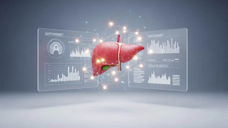
At the technical core of this revolution lies the development of Visualized Transient Elastography, a sophisticated modality that removes the guesswork from traditional tissue stiffness measurements. Historically, technicians often operated in a state of partial blindness, applying mechanical pulses to the liver without a clear real-time view of the specific tissue segment being analyzed. This lack of visual guidance frequently led to sampling errors, especially in patients with complex anatomy or high body mass indices. The current generation of AI-driven systems solves this by overlaying quantitative stiffness maps directly onto high-definition anatomical scans. This synergy allows doctors to precisely target specific regions of interest, ensuring that the data collected is representative of the actual pathological state of the organ. By providing this immediate visual confirmation, the technology increases the confidence of the clinical team and significantly reduces the likelihood of non-diagnostic or misleading results during the examination.
The clinical impact of these integrated visual assessments is most evident in the early detection of liver fibrosis, a stage where medical intervention is most likely to succeed. Research indicates that AI-enhanced ultrasound is exceptionally sensitive to subtle changes in tissue elasticity that precede the visible scarring associated with advanced cirrhosis. Identifying these early-stage markers allows for the implementation of lifestyle changes or pharmacological treatments that can halt or even reverse disease progression. Furthermore, the high negative predictive value of these modern scans is drastically reducing the medical system’s reliance on invasive liver biopsies. While biopsies were long considered the gold standard, they carry risks of hemorrhage and infection, not to mention significant patient discomfort and high costs. By offering a non-invasive alternative that rivals the accuracy of physical tissue samples, these diagnostic platforms are making liver monitoring safer and more tolerable for patients who require long-term surveillance.
Standardizing Results via AI-Powered Automation
A persistent challenge in the field of ultrasound has been the high degree of operator dependency, where the quality of the scan is tethered to the individual skill level of the sonographer. To mitigate this variability, modern systems now incorporate AI-powered co-pilot features that act as an intelligent layer of oversight during the entire diagnostic process. These algorithms are trained on vast datasets of millions of images, allowing them to recognize optimal scanning planes and automatically trigger data acquisition when the best possible view is achieved. This level of automation ensures that the resulting measurements are consistent and standardized, which is an absolute necessity for monitoring a patient’s progress over a multi-year treatment plan. When data is captured according to a uniform set of digital parameters, clinicians can be certain that any changes observed in the liver tissue are the result of physiological shifts rather than technical discrepancies. This reliability transforms the ultrasound into a rigorous, repeatable scientific measurement.
In addition to guiding the capture of images, real-time quality control tools provide instant feedback to the user regarding external variables like probe pressure and patient respiratory patterns. For instance, if a technician applies too much pressure or if a patient’s breathing interferes with the signal, the system provides immediate visual alerts to correct the technique on the fly. This guidance effectively elevates the performance of less experienced practitioners, allowing them to produce results that meet the rigorous standards of expert hepatologists. By democratizing high-quality care in this manner, healthcare facilities can optimize their staffing resources without sacrificing the accuracy of their diagnostic output. As these intelligent systems become more pervasive, the focus of the medical community shifts from the mechanics of image acquisition to the deeper analysis of the data itself. This move toward standardized, AI-assisted diagnostics ensures that every patient receives a world-class assessment, regardless of the specific facility or the individual technician performing the scan.
The Path Forward: Sustaining Diagnostic Excellence
The transition toward an AI-integrated diagnostic model represented a fundamental shift in how the medical community approached chronic liver management and long-term patient outcomes. It became clear that the integration of real-time data analysis and high-resolution imaging served as the definitive solution for the long-standing problem of diagnostic fragmentation and procedural inconsistency. Healthcare administrators who prioritized the adoption of these unified platforms saw immediate improvements in patient throughput and a marked decrease in the need for expensive, invasive follow-up tests. Organizations discovered that investing in AI-driven ultrasound was not merely a hardware upgrade but a strategic move toward a more sustainable and predictive care model. By establishing these high-quality diagnostic benchmarks, clinics were able to provide a level of care that was previously reserved for only the most elite research institutions. This historical shift laid the groundwork for a more equitable healthcare system where precision was accessible to all.
Looking beyond simple detection, the industry successfully moved toward a proactive stance where the diagnostic tool itself provided a roadmap for personalized therapeutic interventions. Medical professionals began to utilize the wealth of quantitative data generated by these systems to create highly specific treatment plans that were tailored to the unique physiological profile of each individual. This shift toward personalized medicine was supported by the seamless integration of ultrasound data into broader digital health records, which allowed for predictive modeling of disease trajectories. As the technology matured, it enabled a collaborative environment where primary care physicians and specialists worked from the same reliable data set, reducing the margin for error and improving communication. The ultimate success of this technological leap was measured not only in clinical accuracy but in the restored quality of life for millions of patients who benefitted from early and accurate intervention. The era of AI-driven liver diagnostics ultimately redefined the boundaries of what was possible in non-invasive medical imaging.