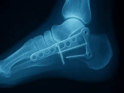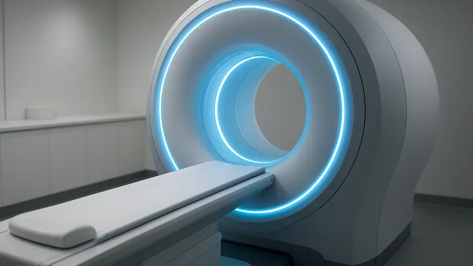

The healthcare sector is currently grappling with a massive diagnostic gap in skeletal health, where millions of adults unknowingly live with brittle bones until a traumatic event forces clinical intervention. The use of deep learning models in medical imaging allows for the precise calculation of bone mass using data that is already being collected for other diagnostic purposes. This innovative approach, often referred to as opportunistic screening, utilizes the thousands of routine computed tomography scans performed daily for reasons unrelated to bone health, such as evaluating abdominal pain or staging cancer. By applying sophisticated algorithms to these existing images, clinicians can identify patients at high risk for fractures without requiring them to undergo additional procedures. This methodology effectively transforms a standard diagnostic tool into a proactive screening platform, potentially catching bone loss years before it results in a debilitating injury and improves long-term outcomes.
The Growing Crisis: Addressing Undiagnosed Bone Loss
Osteoporosis is a systemic skeletal disorder that quietly erodes the micro-architecture of bone tissue, leading to a state where even a minor fall can cause a life-altering fracture. These injuries, particularly in the hip and spine, are not just physical setbacks; they are primary drivers of permanent disability and mortality among the aging population. The difficulty lies in the fact that bone loss is entirely asymptomatic until the moment of structural failure, meaning the majority of individuals living with the condition are unaware of their vulnerability. This silent progression creates a significant burden on the healthcare system, as emergency departments are frequently the first point of contact for patients whose condition should have been managed years earlier. Addressing this crisis requires a fundamental change in how skeletal health is monitored, moving away from reactive treatments and toward comprehensive, early-stage identification strategies implemented before the first fracture occurs.
While the dual-energy X-ray absorptiometry scan serves as the gold standard for bone density measurement, its reach remains remarkably limited in modern clinical practice. Numerous barriers, including a lack of patient awareness, logistical challenges, and low referral rates from primary care physicians, mean that a vast percentage of the at-risk population never undergoes the procedure. Even in regions with robust healthcare infrastructure, the prioritization of other chronic conditions often pushes bone health to the periphery of routine check-ups. Consequently, millions of patients miss the window for early intervention when lifestyle changes or pharmacological treatments are most effective at preserving bone mass. This diagnostic deficit underscores the urgent need for a screening method that integrates seamlessly into existing healthcare workflows, removing the burden of scheduling from the patient while providing physicians with the actionable data they need to initiate timely preventative care.
Deep Learning: Enhancing Bone Analysis Through AI
A research team at the Changzheng Hospital of Naval Medical University recently conducted a retrospective analysis of over five hundred patients to evaluate a new frontier in diagnostic technology. By employing artificial intelligence software built on convolutional neural networks, the researchers demonstrated that it is possible to automate the complex process of bone mineral density assessment. These deep learning tools are trained to recognize the specific contours of the spine, allowing them to perform automatic segmentation of the vertebral bodies with a level of precision that matches human expertise. Once the AI identifies the target vertebrae within a standard CT scan, it can instantly extract volumetric bone density data without any manual intervention from a radiologist. This automation is a key component of the technology’s scalability, as it allows for the analysis of thousands of images in the background of a hospital’s daily operations, turning every compatible scan into a potential screening opportunity.
The technological shift from traditional two-dimensional imaging to three-dimensional volumetric analysis represents a significant leap forward in our understanding of skeletal health. Standard DXA scans provide an areal density measurement, which can sometimes be skewed by the size of the bone or the presence of degenerative changes in the surrounding tissue. In contrast, CT-based AI measures volumetric bone mineral density, offering a three-dimensional perspective that allows for the isolation of specific bone types. Specifically, the software can differentiate between the dense outer cortical shell and the porous inner trabecular bone. This distinction is vital because the trabecular bone is metabolically more active and typically shows the earliest signs of mineral depletion. By focusing on the internal structure of the vertebrae, the AI can detect the initial stages of bone thinning that might be missed by less granular imaging techniques, providing a sensitive early warning system for clinicians.
Measuring Success: Evaluation of Diagnostic Accuracy
To ensure that AI-driven screening is both robust and practical, the Shanghai study tested several measurement strategies involving the lower thoracic and upper lumbar vertebrae. Because routine CT scans are ordered for a wide variety of medical reasons, they do not always capture the same anatomical regions in every patient. For example, a chest scan might only show the upper spine, while an abdominal scan covers the lower regions. By testing four different combinations of the T12, L1, and L2 vertebrae, the researchers aimed to verify if the AI could maintain its accuracy even when only a portion of the spine was visible in the image. This flexibility is essential for real-world application, where the field of view is determined by the primary diagnostic goal rather than the screening objective. The ability to generate reliable data from a variety of scan types ensures that the technology can be applied across multiple hospital departments without requiring the standardization of scanning protocols.
The researchers utilized a rigorous suite of statistical analyses to validate the AI’s performance against the established gold standard of DXA imaging. Through the use of Bland-Altman plots and weighted kappa statistics, the team found exceptional internal consistency across all four measurement strategies, with correlation coefficients reaching as high as 0.909. This high degree of stability indicates that the AI software is capable of producing reproducible results regardless of which specific vertebrae are analyzed. Furthermore, the study examined how well the AI’s categorical diagnoses—sorting patients into normal, osteopenic, or osteoporotic groups—aligned with the results from traditional DXA scans. The findings confirmed a substantial level of concordance, demonstrating that the volumetric data extracted from CT scans is a highly reliable indicator of a patient’s overall skeletal health. Such statistical rigor is necessary to build trust among the medical community, ensuring that AI-generated flags are grounded in sound data.
Clinical Impact: Results From the Shanghai Study
The clinical findings of the research highlighted the impressive precision that modern deep learning models can achieve in a hospital setting. When tasked with identifying patients suffering from osteoporosis, the AI reached a precision rate of approximately 83%. This high level of accuracy means that when the software identifies a patient as having critically low bone density, there is a very high probability that the finding is correct, minimizing the risk of false positives that could lead to unnecessary anxiety. Additionally, the software demonstrated a strong recall rate, successfully identifying nearly three-quarters of all confirmed osteoporosis cases within the patient cohort. These metrics suggest that even though the AI is performing its analysis on scans that were not specifically optimized for bone density, it is still capable of identifying the vast majority of patients who are at risk. This level of performance is more than sufficient for a screening tool designed to trigger further clinical investigation and engagement.
Among the various strategies analyzed during the study, the most comprehensive approach involved averaging the bone mineral density across the T12, L1, and L2 vertebrae. This specific method provided the most stable data set and allowed the algorithm to account for localized variations in bone quality, which can sometimes occur due to minor injuries in a single vertebra. By looking at multiple points along the spine, the AI generates a more holistic view of the patient’s skeletal strength. The research also underscored that this technology is particularly adept at catching cases of osteopenia, the precursor to osteoporosis. Identifying patients during this transitional phase is arguably the most valuable application of the software, as it provides a critical window for intervention before the bone loss becomes irreversible. This capability to detect early-stage degradation highlights the potential for AI to serve as a cornerstone of preventative bone health, moving the needle from crisis management to proactive wellness.
Future Directions: Integrating AI Into Standard Care
While the findings from the research team provide a strong foundation for the use of AI in bone screening, several factors must be addressed to ensure the technology’s effectiveness across diverse populations. The initial study was conducted at a single medical center using specific commercial software, which raises questions about how the algorithm might perform with different CT scanner brands or varying image reconstruction settings. Factors such as the patient’s age and body mass index can also influence the appearance of bone tissue on a scan, potentially affecting the AI’s density calculations. To achieve universal reliability, the software must be validated across a wider range of clinical environments and patient demographics. Researchers have pointed out that while the AI is an exceptional tool for identification, its current role is best suited as a secondary screening mechanism. It is designed to work in tandem with traditional methods, acting as a high-tech sieve that catches potential cases and routes them toward confirmation.
The study successfully established that AI-driven volumetric analysis of routine CT scans was a powerful and viable method for uncovering hidden bone loss. By providing a precise and automated way to monitor the health of the T12 to L2 vertebrae, the technology proved its ability to function as a reliable early warning system. Health systems that integrated these tools began to see a more streamlined path from incidental findings to active patient management, ensuring that skeletal health was no longer overlooked. The transition from reactive to proactive care was bolstered by the realization that valuable diagnostic data was already hidden within existing medical records. Moving forward, the clinical community looked to expand these AI applications beyond the spine, exploring other skeletal regions to create a more complete picture of fracture risk. By making bone density screening a standard byproduct of routine imaging, medical professionals took a decisive step toward eliminating the silent epidemic of osteoporosis.