
A groundbreaking multicenter study has demonstrated that artificial intelligence can now analyze internal tumor architectures to identify high-risk biological signatures non-invasively. This advancement addresses a long-standing obstacle in the treatment of hepatocellular carcinoma, where clinicians often operate without a full understanding of a tumor’s underlying aggressiveness. For decades, the standard of care for liver cancer has relied heavily on surgical resection, a process that physically removes the mass but cannot always account for the microscopic variables that dictate long-term success. Currently, surgeons face a persistent diagnostic gap because the most reliable indicators of a high-risk tumor are typically only revealed after the tissue has been removed and examined by a pathologist. This retrospective approach creates a high-stakes scenario where the true danger of the disease is identified only after the primary window for personalized intervention has closed, leaving patients and doctors to react to the cancer’s return rather than preventing it through early, tailored strategies.
Decoding Tumor Aggressiveness Through Digital Mapping
Defining High-Risk Markers: The Need for Non-Invasive Tools
The primary objective of this sophisticated computational approach is the accurate prediction of the MSE score, a clinical metric that defines high-risk hepatocellular carcinoma based on several critical pathological features. These features include microvascular or macrovascular invasion, where cancer cells infiltrate blood vessels to spread elsewhere, as well as the presence of satellite nodules—small secondary tumors that hide in the surrounding liver tissue. Furthermore, a high Edmondson-Steiner grade indicates that the cancer cells have mutated significantly away from healthy tissue, signaling a highly aggressive and unpredictable trajectory. Because these markers are microscopic or situated deep within the organ’s structure, they remain largely invisible during standard preoperative imaging. Identifying these specific traits is essential for determining which patients are at a higher risk of early recurrence, but the industry has historically lacked the precision tools required to make such determinations without invasive surgical intervention or post-operative analysis.
While a traditional physical biopsy could theoretically identify these aggressive traits, the procedure is fraught with clinical risks that often outweigh its diagnostic benefits in liver cancer cases. Inserting a needle into a vascular tumor like a hepatocellular carcinoma carries a substantial risk of internal bleeding and the terrifying possibility of tumor seeding, where cancer cells are inadvertently dragged along the needle track and spread into previously healthy areas. Consequently, the medical community has sought a “virtual biopsy” that can extract the same biological insights through advanced imaging alone. By leveraging artificial intelligence to interpret complex data patterns from standard scans, researchers are now providing a safer alternative that protects patient health while delivering high-level diagnostic clarity. This shift toward non-invasive risk assessment represents a significant leap forward in patient safety, as it allows for the creation of a comprehensive biological profile before a single incision is made, ensuring that the surgical plan is as informed as possible from the very start.
Technical Foundations: Habitat Imaging and Contrast Analysis
The technical success of this predictive model relies on the use of a specialized contrast agent known as gadobenate dimeglumine, or Gd-BOPTA. Unlike common contrast agents that merely follow the flow of blood, Gd-BOPTA is partially absorbed by healthy liver cells and eventually excreted into the bile ducts. This unique property allows for a “hepatobiliary phase” in MRI scans, where healthy liver tissue glows brightly while the cancerous tumor appears as a dark, distinct void. This sharp contrast provides an exceptionally detailed look at the interface where the cancer meets the surrounding liver, revealing subtle irregularities in the tumor’s border and the health of the adjacent tissue. By focusing on this critical boundary, the AI can detect signs of infiltration that the human eye might overlook. This imaging technique serves as the raw data source for the machine learning algorithms, providing the high-resolution evidence needed to differentiate between localized masses and those that have already begun to compromise the surrounding anatomy.
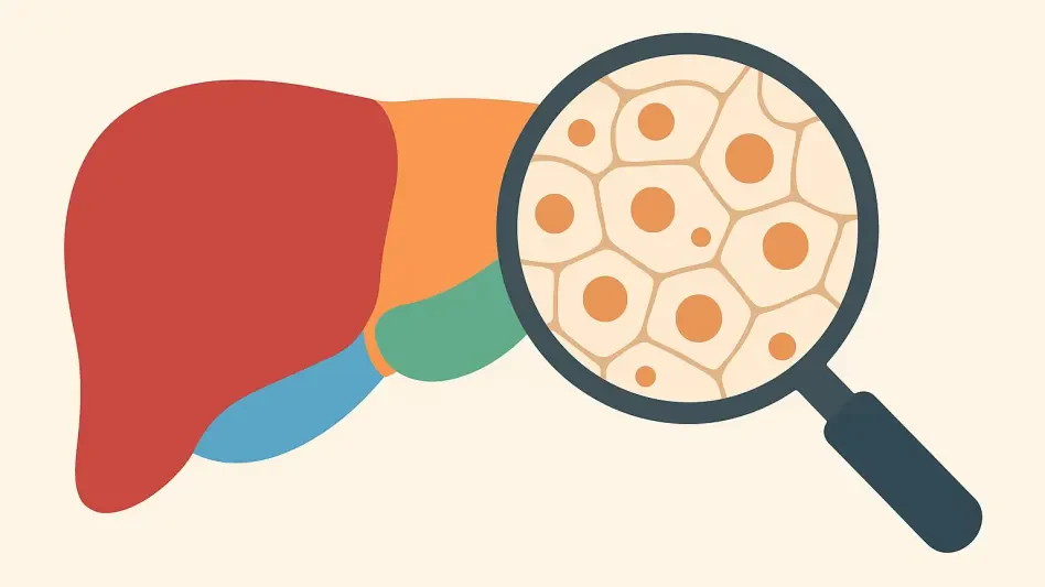
A core innovation in this research is the application of “habitat imaging,” a concept that treats a tumor not as a uniform lump of tissue, but as a complex and diverse ecosystem. Using a machine-learning algorithm called k-means clustering, the artificial intelligence divides the tumor into various “habitats” or subregions based on their specific blood flow patterns and cellular density. By analyzing ratio maps that compare the arterial phase—when blood first enters the tumor—with the hepatobiliary phase, the AI identifies unique fingerprints of aggressiveness within the mass. These subregions may include necrotic cores where tissue has died, or highly active cellular zones that suggest rapid growth. The spatial patterns, textures, and intensity distributions of these internal habitats contain hidden information about the tumor’s biological behavior. This granular level of analysis allows clinicians to see the internal architecture of the cancer, turning a static image into a dynamic map of the disease’s potential to grow, spread, and eventually recur after surgical treatment.
Evaluating Clinical Performance and Surgical Impact
The Five Pillars: Preoperative Risk Prediction
Through the analysis of hundreds of patients across multiple medical institutions, the research team identified five independent factors that serve as the foundation for their predictive model. These factors include blood-based markers like Alpha-Fetoprotein (AFP) levels, which have long been associated with liver cancer activity, and specific MRI patterns such as corona enhancement and peritumoral hypointensity. Corona enhancement refers to a ring of contrast around the tumor that often signals microscopic invasion at the edges, while peritumoral hypointensity suggests that the surrounding liver tissue is being actively compromised by the malignancy. Interestingly, the study also identified that “non-rim arterial phase hyperenhancement,” a classic appearance for many tumors, actually served as a protective factor in this context. This finding suggests that tumors following a more “typical” growth pattern might actually be less aggressive than those exhibiting unusual or irregular enhancement characteristics, providing a nuanced view of cancer pathology.
The most influential component of this predictive model was the Habitat Score, which emerged as a dominant indicator of patient risk. With an impressive odds ratio of 44.46, the AI-derived score for the tumor’s internal organization revealed that patients with unfavorable habitat signatures were over forty-four times more likely to harbor high-risk disease than those with more uniform tumor structures. To make this complex data accessible to clinicians, these five variables were integrated into a “nomogram”—a graphical tool that allows doctors to input specific patient data and receive a clear, actionable probability score for high-risk disease. This tool bridges the gap between high-level data science and everyday medical practice, giving surgeons a reliable way to quantify risk before they enter the operating room. By providing a single, consolidated score, the nomogram empowers medical teams to move away from subjective assessments and toward a data-driven approach that accounts for the specific biological reality of each individual patient’s condition.
Validation of Results: Measuring Long-Term Outcomes
For any artificial intelligence model to be effective in a clinical setting, it must demonstrate generalizability, meaning it can perform accurately across different hospitals and with various types of imaging equipment. The multicenter study from China rigorously tested this model, achieving an initial Area Under the Curve (AUC) of 0.844 during the training phase. When the model was subsequently applied to an external group of patients from three different hospitals, it maintained a robust AUC of 0.820. In the field of medical diagnostics, a score above 0.80 is widely considered to be a strong indicator of a reliable and clinically useful tool. This consistency across multiple institutions suggests that the AI is not just “learning” the quirks of one specific data set, but is instead identifying universal biological truths about how aggressive liver cancer presents on an MRI. This validation is a critical step in ensuring that the technology can be deployed widely to benefit patients regardless of where they receive their care.
The practical value of the AI model was further confirmed by its ability to predict long-term recurrence-free survival (RFS), which is the primary concern for any patient undergoing cancer surgery. The researchers found a dramatic difference in outcomes between patients flagged as high-risk and those identified as low-risk. Patients with high-risk habitat signatures had a median cancer-free period of only 18 to 19 months, whereas low-risk patients remained free of the disease for a median of 56 to 69 months. This stark contrast confirms that the AI is accurately detecting the fundamental biological clock of the cancer, rather than just identifying arbitrary shapes on a scan. By revealing which patients are likely to face an early recurrence, the model provides a window of opportunity for doctors to intervene sooner and more aggressively. This predictive power allows for a shift in the oncology timeline, moving the focus from post-operative crisis management to a proactive, long-term strategy that prioritizes the most vulnerable patients from the moment of diagnosis.
Reshaping Strategy: The Path Toward Precision Oncology
The ability to identify high-risk hepatocellular carcinoma before an operation fundamentally alters the surgical landscape, allowing for a level of customization that was previously impossible. If a surgeon knows a tumor is highly aggressive before the first incision, they can modify their technique to include wider surgical margins, removing more of the surrounding “healthy” tissue to ensure that no microscopic satellite nodules are left behind. Furthermore, patients with high-risk scores can be prioritized for neoadjuvant therapies—such as targeted drug regimens or localized radiation—designed to shrink the tumor and reduce its biological volatility before surgery is even attempted. In some instances, a high-risk AI assessment might even shift a patient’s treatment path entirely, moving them higher up the list for a liver transplant, which remains the most effective cure for aggressive or multifocal disease. These adjustments ensure that the intensity of the treatment matches the intensity of the cancer.
As the medical community continues to integrate AI-driven habitat imaging into standard protocols, the focus remains on refining these tools for even greater accuracy and broader application. The study concluded that the fusion of advanced contrast imaging with machine learning successfully provided a non-invasive alternative to traditional pathology. Moving forward, the goal should be to incorporate these predictive scores into routine clinical workflows and to conduct larger, prospective trials that can further validate the impact on overall survival rates. For practitioners, the immediate takeaway is the necessity of looking beyond the surface of a tumor to understand its internal “geography.” For patients, this technology represents a significant step toward a future where treatment is not a gamble, but a precisely calculated response to the specific biological threats they face. The transition to this level of precision oncology will likely define the next decade of liver cancer care, turning hidden biological risks into visible, manageable data points that save lives.