
The shift from traditional analog systems to high-precision digital mammography has fundamentally altered how medical professionals identify malignancies before they become life-threatening. The integration of state-of-the-art imaging systems at institutions like Orillia Soldiers’ Memorial Hospital represents a significant advancement in the healthcare sector. This review explores the evolution of the technology, its key features, performance metrics, and the impact it has had on breast cancer screening and patient care. The purpose of this review is to provide a thorough understanding of the technology, its current capabilities, and its potential future development in early disease detection.
Evolution and Core Principles of Modern Breast Imaging
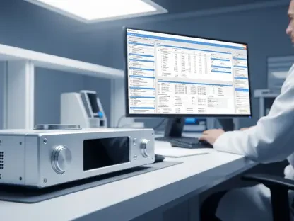
Modern breast imaging has transitioned from basic film-based processes to high-resolution digital capture, emphasizing the need to replace aging, end-of-life equipment. These new systems operate on the principle of direct radiography, where X-ray energy is converted into a digital signal almost instantaneously. This method eliminates the degradation of detail often associated with older physical processing techniques.
This technological evolution is highly relevant in the broader healthcare landscape where early intervention is the primary driver of improved patient outcomes. By providing a digital-first approach, medical centers can ensure that imaging data is easily stored, shared, and analyzed. Such modernization is not merely a convenience; it is a fundamental requirement for maintaining diagnostic standards as legacy systems become obsolete and difficult to maintain.
Critical Technical Features and Design Enhancements
High-Resolution Diagnostic Clarity
Modern mammography units provide clearer and crisper images, allowing radiologists to better distinguish potential abnormalities from healthy tissue. This high-resolution output is achieved through advanced sensor technology that captures minute variations in tissue density. The increased clarity is vital for spotting calcifications or small masses that might be obscured in lower-quality scans.
Increased diagnostic confidence directly impacts the clinical workflow by reducing false positives and improving the accuracy of early-stage intervention. When radiologists have access to superior imaging, the need for stressful follow-up biopsies often decreases. This precision ensures that medical resources are directed toward patients who truly require urgent care, streamlining the entire diagnostic process.
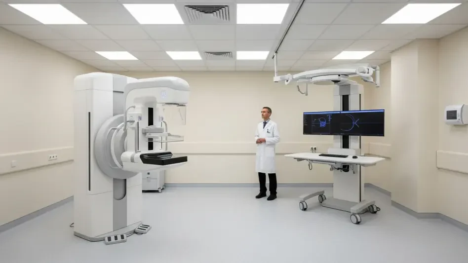
Human-Centric Ergonomics and Patient Comfort
Physical design in modern equipment addresses the psychological and physical stress often associated with cancer screenings. New machines feature rounded edges, warmer surfaces, and refined compression paddles that adapt to the patient’s anatomy. These design choices are intended to mitigate the discomfort that frequently leads to screening avoidance among high-risk populations.
The technical aspects of pressure regulation and positioning play a crucial role in putting patients at ease during the imaging process. Sensors now monitor the exact amount of force applied, ensuring optimal image quality without unnecessary pain. By prioritizing the patient experience, manufacturers are helping to increase compliance with regular screening intervals, which is essential for community-wide health.
Emerging Trends and Technological Innovations
The field is currently moving toward contrast-enhanced mammography and the integration of digital tools to streamline clinical workflows. Contrast-enhanced techniques allow for a better view of blood flow patterns, which can highlight aggressive tumors that might otherwise remain hidden. This shift moves the industry beyond traditional 2D imaging, offering a more functional perspective on breast health.
These innovations influence the trajectory of preventative medicine by providing a more personalized diagnostic experience. Digital integration also allows for the use of advanced algorithms that assist in triaging cases based on urgency. As these tools become more sophisticated, the focus shifts from simple observation to a proactive, data-driven analysis of patient risk factors.
Real-World Applications and Community Implementation
The deployment of this technology at the Orillia Soldiers’ Memorial Hospital Diagnostic Imaging department serves as a primary example of regional implementation. By installing high-performance equipment locally, the facility ensures that residents do not have to travel to major urban centers for life-saving screenings. This decentralization of advanced care is a critical factor in improving community health accessibility.
Providing high-quality medical services within the local community fosters a stronger relationship between patients and the healthcare system. When a hospital modernizes its infrastructure, it sends a clear signal of commitment to the well-being of its population. This regional accessibility reduces the logistical barriers to healthcare, ensuring that more individuals receive timely diagnostic services.
Navigating Technical and Financial Challenges
A significant challenge facing this technology is the high cost of acquisition and the subsequent reliance on community-led philanthropy to modernize infrastructure. Many hospitals find that government funding alone is insufficient to keep pace with the rapid cycle of technical obsolescence. Consequently, local donations become the lifeblood of diagnostic innovation, creating a variable landscape of care based on regional fundraising capacity.
Ongoing development efforts aim to mitigate technical hurdles related to equipment maintenance and the transition from legacy systems. Sophisticated hardware requires specialized technicians and constant software updates to remain secure and effective. Navigating these financial and technical complexities requires a long-term strategic vision that balances the need for innovation with the realities of budgetary constraints.
Future Outlook for Mammography Infrastructure
The current state of mammography technology serves as a foundation for future breakthroughs in imaging precision. As digital platforms become more versatile, they will likely integrate more seamlessly with artificial intelligence to provide real-time diagnostic support. This evolution will move the needle from reactive screening to a more predictive model of breast health management.
Sustainable and versatile diagnostic tools will have a long-term impact on the broader medical industry and preventative care. The goal is to create an infrastructure where equipment can be upgraded modularly rather than being replaced entirely. This approach will ensure that healthcare facilities can remain at the cutting edge of science without the prohibitive costs associated with full-scale equipment turnover.
Final Assessment of Diagnostic Advancements
The review of advanced mammography systems demonstrated a significant synergy between technical precision and patient-centered care. These systems successfully addressed the limitations of legacy equipment by providing superior image quality and improved ergonomic features. The integration of such technology at the regional level proved to be a vital step in modernizing local healthcare and ensuring equitable access to high-standard diagnostics.
The transition to high-resolution digital systems established a new standard for early disease detection, significantly improving the chances of successful treatment. Community involvement and philanthropic support emerged as essential components in the successful deployment of these medical advancements. Ultimately, the focus on technical clarity and patient comfort ensured that the diagnostic process became both more effective and less intimidating for the general public.