The medical field is on the brink of a significant breakthrough with the development of a new lung scanning method. This innovative technique, pioneered by researchers from Newcastle University in the UK, utilizes a specialized gas visible on MRI scans to provide real-time insights into lung function. This advancement is poised to transform the treatment and monitoring of respiratory diseases, offering new hope to patients with conditions such as asthma, Chronic Obstructive Pulmonary Disease (COPD), and post-lung transplant complications.
Innovative Lung Scanning Method
Development and Functionality
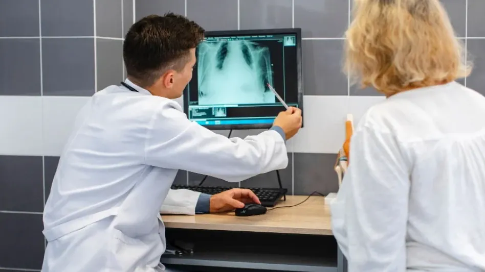
Researchers at Newcastle University have developed a groundbreaking lung scanning method using perfluoropropane gas. This gas is safe for patients to inhale and is visible on MRI scans, allowing doctors to observe how air flows in and out of the lungs. The ability to visualize poorly ventilated areas in real-time provides a detailed understanding of lung function, which is crucial for diagnosing and treating respiratory diseases.
With this innovative technique, doctors can now observe lung performance as it happens, identifying specific areas of concern immediately. Traditional methods, which often involved delayed results and less accurate imaging, are now being overshadowed by this real-time scanning capability. The discovery and successful implementation of this method could lead to significant advancements in providing timely and effective treatments.
Mapping Ventilation Patterns
The new scanning technique is particularly effective in identifying areas of the lung with patchy ventilation. By mapping these ventilation patterns, medical professionals can pinpoint specific regions with ventilation defects. This detailed insight is essential for personalizing care and implementing early interventions, ultimately improving patient outcomes.
Through detailed imaging, healthcare providers can see exactly which areas of the lung are not receiving adequate air, allowing them to target treatment precisely where it is needed. This ability to map out the lung’s ventilation in such detail is a groundbreaking step forward. In cases where asthma or COPD causes uneven air distribution in the lungs, the real-time map generated by this technique becomes an invaluable tool for both diagnosis and ongoing treatment management.
Clinical Applications in Asthma and COPD
Initial Clinical Trials
Initial clinical trials have demonstrated the efficacy of this lung scanning method in patients with asthma and COPD. By measuring ventilation improvement after treatment with bronchodilators such as salbutamol, researchers can quantify the degree of improvement and assess the effectiveness of treatments. This capability is invaluable for clinical trials of new lung disease treatments, providing a reliable method for evaluating therapeutic outcomes.
In these early trials, patients undergoing treatment were monitored using the new scanning method, allowing researchers to see how their lungs responded in real-time. This data is crucial not only for assessing individual patient progress but also for gauging the overall effectiveness of new treatments being tested. The real-time feedback provided by the scans helps fine-tune therapeutic interventions, making them more effective in managing respiratory conditions.
Personalized Treatment Plans
The ability to visualize lung function in real-time allows for the development of personalized treatment plans. By identifying poorly ventilated areas, doctors can tailor treatments to address specific needs, optimizing therapeutic outcomes for each patient. This personalized approach is a significant advancement in the management of respiratory diseases, offering new hope for improved quality of life.
Patients can benefit from treatments that are specifically designed to target their unique condition and lung function patterns. This move towards more personalized medical interventions means that treatments can be more effective, reducing unnecessary medication and focusing on what will truly help. Personalized treatment plans based on real-time data represent a paradigm shift in how respiratory diseases are managed, leading to better health outcomes and enhanced quality of life for sufferers.
Monitoring Lung Transplant Recipients
Real-Time Lung Function Measurements
One of the most promising applications of this new scanning method is in monitoring lung transplant recipients. The ability to provide real-time lung function measurements allows for early detection of complications such as chronic lung allograft dysfunction. This condition, often caused by an immune response against donor lungs, can be detected early, enabling timely intervention and potentially preventing further damage.
For lung transplant recipients, this early detection is a crucial advantage. The ability to monitor the functionality of transplanted lungs in real-time means that doctors can spot issues before they become severe. This proactive approach can help prevent the failure of the transplant, which is often a risk due to the body’s immune response. By catching these signs early, treatment can be adjusted quickly to maintain the health of the transplanted lungs.
Early Detection and Intervention
Early detection of lung function decline is crucial for lung transplant recipients. The new scanning method offers a dynamic viewpoint of the lung’s response to treatments, allowing for prompt adaptation and personalized medical care. This capability is essential for protecting transplanted lungs and improving patient outcomes, making it a significant advancement in post-transplant care.
Patients receiving lung transplants can face numerous challenges, including the risk of chronic rejection and other complications. The ability to see real-time lung function means that any deviations from expected recovery can be addressed immediately. This dynamic monitoring approach not only safeguards the health of transplanted lungs but also ensures that recipients have the highest possible chance of long-term success. This breakthrough provides a much-needed safety net for transplant recipients, leading to significantly better health outcomes.
Broader Implications and Future Directions
Impact on Respiratory Disease Treatment
The introduction of this innovative lung scanning method marks a significant advancement in respiratory disease treatment. The ability to monitor lung function in real-time, detect early signs of decline, and personalize treatments has the potential to revolutionize the management of respiratory diseases. This breakthrough offers new hope for patients and sets a precedent for future research and development in the field.
The broad implications of this technology extend beyond initial use cases. Its application could benefit various respiratory conditions, leading to early diagnosis and appropriate intervention for a range of diseases. The shift towards more individualized treatment plans is set to change the landscape of respiratory healthcare, ensuring that each patient receives care best suited to their condition.
Collaborative Research and Development
The medical community is on the cusp of a groundbreaking development with a new method for lung scanning. Researchers at Newcastle University in the UK have developed this cutting-edge technique, which employs a special gas visible through MRI scans to provide real-time data on lung function. This revolutionary approach is likely to change how respiratory diseases like asthma, Chronic Obstructive Pulmonary Disease (COPD), and complications following lung transplants are treated and monitored.
The new scanning method is a significant improvement over traditional imaging techniques, which often fail to offer the same level of detailed insights into the functioning of the lungs. By using this specialized gas, doctors can observe the behavior of the lungs more accurately than ever before. This real-time analysis could lead to earlier diagnosis, better treatment plans, and improved outcomes for patients dealing with chronic respiratory conditions. As the technique becomes more widespread, it is expected to bring a much-needed ray of hope to those struggling with severe lung-related health issues, significantly enhancing their quality of life.
