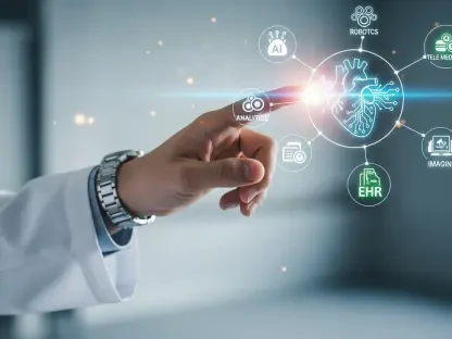

Peering into a medical scan reveals far more than the human eye can perceive; within each pixel lies a wealth of untapped data that holds the key to predicting disease behavior and personalizing patient treatment. Radiomics represents a significant advancement in quantitative medical imaging and personalized medicine. This review will explore the evolution of this technology, its core workflow, key predictive modeling techniques, and its impact on oncological and non-oncological applications. The purpose of this review is to provide a thorough understanding of radiomic biomarker prediction, its current capabilities, and its potential future development.
Fundamentals of Radiomics and Quantitative Imaging
At its core, radiomics serves as a crucial bridge connecting macroscopic medical imaging with microscopic molecular biology. This field operates on the principle of converting standard clinical images—such as those from Computed Tomography (CT), Magnetic Resonance Imaging (MRI), or Positron Emission Tomography (PET)—into high-dimensional, mineable data. By systematically extracting hundreds or even thousands of quantitative features, radiomics creates sophisticated digital biomarkers that can characterize disease, forecast patient outcomes, and predict responses to specific therapies.
This quantitative approach stands in stark contrast to traditional radiological assessment, which has historically relied on qualitative interpretation and visual inspection. While invaluable, human interpretation is inherently subjective and limited in its ability to detect the subtle, complex patterns of texture and shape that signify underlying biological processes. Radiomics augments this clinical expertise by providing an objective, data-driven layer of analysis, positioning itself as an essential tool in the broader landscape of precision medicine, where treatment is tailored to the individual patient.
The Radiomic Workflow: From Image to Prediction
The journey from a raw medical image to a clinically actionable prediction follows a structured, multi-step process. Each stage of the radiomic workflow is critical, with technical decisions made early on having a profound impact on the final model’s accuracy, robustness, and reproducibility. This meticulous pipeline ensures that the information extracted is both meaningful and reliable.
Medical Image Acquisition and Pre-processing
The foundation of any radiomic analysis is the quality and consistency of the medical images themselves. Data acquisition involves various imaging modalities, each with its own set of parameters, such as scanner vendor, reconstruction algorithms, and contrast protocols. Variations in these settings can introduce significant biases that obscure true biological signals. Consequently, standardization of acquisition protocols is paramount for developing models that can be generalized across different hospitals and patient populations.
To mitigate these inconsistencies, rigorous pre-processing steps are essential. Techniques such as image normalization, which standardizes pixel intensity values, and artifact correction, which removes noise or distortions, are applied to harmonize the data. These foundational procedures ensure that the extracted radiomic features reflect the underlying pathophysiology of the tissue rather than technical variability, thereby enhancing the reliability of any downstream analysis and predictive modeling.
Segmentation and Region of Interest Definition
Once an image is acquired and pre-processed, the next critical step is to precisely delineate the region of interest (ROI), such as a tumor, lesion, or specific organ. This process, known as segmentation, isolates the anatomical area from which features will be extracted. Historically, segmentation has been a laborious manual task performed by radiologists, a method that is both time-consuming and subject to inter-observer variability.
To address these limitations, semi-automated and fully automated segmentation methods have gained prominence. Semi-automated tools often require minimal user input to refine boundaries, improving efficiency. More recently, fully automated approaches leveraging artificial intelligence, particularly deep learning models like U-Nets, have demonstrated the ability to perform segmentation with accuracy rivaling or even exceeding human experts. The choice of segmentation method directly influences the stability of the extracted features and, ultimately, the clinical viability of the final predictive model.
Feature Extraction and Quantification
The heart of the radiomic process is the automated extraction of a vast array of quantitative features from the segmented ROI. These features are designed to capture distinct characteristics of the tissue’s phenotype, providing a comprehensive digital signature of the disease. They are typically categorized into several classes, each offering unique insights.
First-order features describe the distribution of voxel intensities within the ROI, akin to a histogram, quantifying aspects like mean intensity, skewness, and kurtosis. Second-order, or texture, features analyze the spatial relationships between voxels, capturing patterns of tissue heterogeneity using metrics derived from matrices like the gray-level co-occurrence matrix. Higher-order features involve applying filters or mathematical transforms, such as wavelets or Laplacian of Gaussian filters, to the image before quantification, enabling the analysis of patterns at different scales and textures that are not visually apparent.
Predictive Modeling and Signature Development
With a high-dimensional feature set in hand, the final step involves using machine learning to build a predictive model. Given that the number of features often far exceeds the number of patients in a study, a key challenge is to select the most relevant and stable features to avoid overfitting. Techniques like LASSO regression or recursive feature elimination are commonly used for this purpose.
Once an optimal feature subset is identified, various modeling algorithms can be employed. Classic machine learning models, such as Random Forests, Support Vector Machines (SVMs), and logistic regression, are widely used for their interpretability and effectiveness with structured data. The result of this process is a “radiomic signature”—a specific combination of features and their associated weights that generates a predictive score. This signature is then rigorously validated on independent datasets to confirm its performance before it can be considered for clinical use.
Innovations and Evolving Methodologies
The field of radiomics is in a constant state of evolution, driven by advancements in artificial intelligence and a deeper understanding of molecular biology. These innovations are pushing the boundaries of what is possible, enabling more sophisticated analyses and yielding deeper biological insights from medical images.
Deep Learning and End-to-End Radiomics
The rise of deep learning, particularly convolutional neural networks (CNNs), has introduced a paradigm shift in radiomics. Traditional radiomics relies on pre-defined, “engineered” features. In contrast, deep learning models can perform “end-to-end” analysis, learning relevant feature representations directly from the raw image data without explicit programming. This approach allows the model to identify novel, complex patterns that may not be captured by conventional feature sets.
These deep learning-based radiomic models have shown immense promise in various predictive tasks. By bypassing manual feature extraction and selection, they can potentially create more robust and powerful biomarkers. However, this comes with the trade-off of reduced interpretability, as the features learned by the network are often abstract and not easily understood, a challenge often referred to as the “black box” problem.
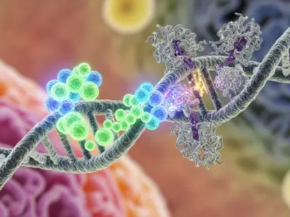
Radiogenomics: Linking Imaging to Molecular Data
Radiogenomics represents a particularly exciting frontier, aiming to establish a direct link between imaging phenotypes and underlying molecular data. This integrative field investigates the correlation between radiomic features and genomic information, such as gene mutations, expression levels, and molecular pathways. By doing so, it provides a non-invasive window into the tumor’s biological makeup.
This integration allows for the prediction of crucial molecular biomarkers directly from standard-of-care scans. For example, radiogenomic models can forecast the presence of an EGFR mutation in lung cancer or ID# status in gliomas, information that is critical for guiding targeted therapies. This capability to infer molecular characteristics without an invasive biopsy holds the potential to revolutionize personalized medicine, making it faster, safer, and more accessible.
Clinical Applications in Biomarker Prediction
The theoretical promise of radiomics is increasingly being realized in clinical practice, with a growing number of applications demonstrating its value as a diagnostic, prognostic, and predictive tool across diverse medical specialties.
Oncology Diagnosis: Prognosis and Treatment Response
Oncology is the field where radiomics has seen its most mature development and application. In non-small cell lung cancer, radiomic models built from CT scans have successfully predicted EGFR mutation status, helping to guide decisions on targeted therapy. Similarly, in melanoma, features from PET/CT images are being used to forecast a patient’s response to immunotherapy, allowing clinicians to identify likely responders and non-responders early in the treatment course.
Beyond treatment response, radiomics is also used for prognosis and risk stratification. In prostate cancer, MRI-based radiomic signatures can help distinguish between indolent and aggressive tumors, aiding in decisions about active surveillance versus immediate intervention. Likewise, in breast cancer, radiomic features have shown potential in predicting the risk of recurrence, providing valuable information for tailoring post-treatment follow-up strategies.
Neurology and Cardiology
The utility of radiomics extends far beyond cancer care. In neurology, researchers are using radiomic features extracted from brain MRIs to predict the rate of cognitive decline in patients with Alzheimer’s disease and to differentiate between various types of dementia. This provides a quantitative tool to monitor disease progression and assess the efficacy of new therapeutic interventions.
In cardiology, radiomics is being applied to characterize cardiovascular diseases non-invasively. For instance, features derived from coronary CT angiography can help assess the vulnerability of atherosclerotic plaques, identifying those at high risk of rupture that could lead to a heart attack. Furthermore, cardiac MRI radiomics is emerging as a powerful method for quantifying myocardial fibrosis and inflammation, which are key indicators of heart failure and other cardiac conditions.
Challenges and Hurdles to Clinical Translation
Despite its immense potential, the path for radiomics from a research tool to a staple of routine clinical practice is fraught with significant challenges. Overcoming these technical and regulatory hurdles is essential for realizing the full promise of this technology.
The Reproducibility and Standardization Crisis
One of the most significant obstacles to the widespread adoption of radiomics is the issue of reproducibility. Radiomic features can be highly sensitive to variations in image acquisition parameters, scanner hardware, and reconstruction settings. A model developed at one institution may not perform well at another if imaging protocols differ, leading to a “reproducibility crisis.”
To combat this, global efforts like the Image Biomarker Standardization Initiative (IBSI) are working to harmonize feature definitions and calculation methods. These initiatives aim to create a common language for radiomics, ensuring that features are computed consistently regardless of the software used. Achieving this level of standardization is a critical prerequisite for building robust and generalizable models that can be trusted in a clinical setting.
The Need for Prospective Validation and Regulatory Approval
The vast majority of radiomic studies published to date have been retrospective in nature, meaning they were developed and tested on existing datasets. While these studies are valuable for hypothesis generation, they do not provide the high level of evidence required for clinical implementation. To prove its true clinical utility, a radiomic signature must be validated in large-scale, multi-center prospective clinical trials.
Furthermore, for a radiomic model to be used in clinical decision-making, it often needs to navigate a complex regulatory landscape. Depending on its intended use, a radiomic tool may be classified as a medical device, requiring approval from bodies like the U.S. Food and Drug Administration (FDA) or the European Medicines Agency (EMA). This process is rigorous and requires extensive evidence of the model’s safety, accuracy, and clinical benefit.
Future Outlook and Long Term Impact
Looking ahead, the trajectory of radiomics points toward deeper integration into the fabric of modern medicine. The continued convergence of imaging technology, artificial intelligence, and big data analytics will likely unlock even more powerful applications. In the near future, we can expect to see radiomic tools seamlessly integrated into clinical decision support systems, providing clinicians with real-time, quantitative insights directly within their existing workflows.
The long-term impact of radiomics could be transformative. By enabling real-time analysis of disease at a level of detail previously unimaginable, it has the potential to shift the paradigm of healthcare from reactive to proactive. It will play a pivotal role in the early detection of disease, the precise monitoring of treatment response, and the development of truly personalized therapeutic strategies, ultimately shaping a future of more efficient, effective, and data-driven healthcare for all.
Conclusion: A Synthesis of the Field
This review charted the landscape of radiomic biomarker prediction, from its foundational principles to its advanced clinical applications. The journey through the meticulous workflow—encompassing image acquisition, segmentation, feature extraction, and machine learning—highlighted the technology’s capacity to convert standard medical scans into powerful predictive instruments. The analysis of evolving methodologies, particularly deep learning and radiogenomics, revealed a field rapidly advancing toward more profound biological insights. While significant hurdles related to standardization and clinical validation remained, the demonstrated impact across oncology, neurology, and cardiology underscored its transformative potential. Ultimately, radiomics established itself not as a futuristic concept but as a cornerstone technology poised to redefine the future of diagnostic medicine and personalized treatment.