
Managing a pregnant patient who presents with sudden and severe abdominal pain involves a high-stakes clinical balancing act that requires physicians to weigh the immediate necessity of an accurate diagnosis against the long-term safety of the developing fetus. This complex scenario often puts medical professionals in a difficult position where they must choose between high-clarity imaging tools and the imperative to protect both the mother and the unborn child from ionizing radiation or invasive procedures. The challenge is further intensified because pregnancy itself frequently alters baseline biological metrics, such as heart rate and white blood cell counts, which can easily mask the onset of serious infections or inflammatory conditions. When a uterus expands to accommodate growth, it physically shifts the location of internal organs, making traditional physical examinations less reliable than in non-pregnant patients. Consequently, diagnostic hesitancy can lead to dangerous delays, increasing the risk of life-threatening complications like organ perforation or localized abscesses.
Evaluating Imaging Modalities for Fetal Safety
Limitations of Ultrasound and Computed Tomography
Ultrasound remains the frontline diagnostic tool for investigating abdominal pain in expectant mothers primarily because it is cost-effective, portable, and completely free of ionizing radiation. While it excels in early pregnancy, its diagnostic utility often declines significantly as the patient moves into the second and third trimesters. This decline occurs because the increasing size of the fetus and the expansion of the uterus create significant physical barriers, often referred to by radiologists as acoustic shadows, which obscure deeper abdominal structures. Furthermore, the presence of bowel gas and the repositioning of the appendix can make it nearly impossible to visualize specific areas of concern with the necessary precision. In many complex clinical presentations, ultrasound provides inconclusive results, which necessitates the use of more advanced imaging to prevent a missed diagnosis that could result in adverse outcomes for the pregnancy.
Building on this foundation, Computed Tomography (CT) is frequently considered the gold standard for diagnosing acute abdominal issues in the general population due to its high resolution and speed. However, its application during pregnancy is strictly managed under rigorous protocols because the ionizing radiation it emits poses potential risks to fetal cellular development. While modern CT technology allows for lower radiation doses, medical teams generally reserve this modality for life-threatening trauma situations where the survival of the mother is the primary clinical objective and rapid imaging is mandatory. Additionally, the use of intravenous contrast agents in CT scans introduces another layer of complexity, as these substances are typically avoided unless other methods have failed to clarify the clinical picture. The decision to use CT must always be a carefully weighed trade-off, ensuring that the diagnostic benefit clearly outweighs the potential risks associated with radiation exposure.
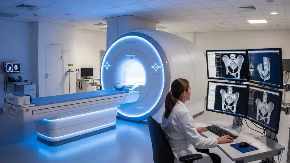
The Clinical Efficacy of Magnetic Resonance Imaging
Magnetic Resonance Imaging (MRI) has increasingly become a cornerstone of maternal-fetal medicine because it provides high-resolution anatomical detail without the biological risks associated with ionizing radiation. One of the most significant technological advancements in this field is the implementation of Diffusion-Weighted Imaging (DWI), a specific sequence that allows radiologists to detect inflammation and infection by monitoring the microscopic movement of water molecules within bodily tissues. Because this technique does not require the administration of contrast dyes to visualize pathology, it significantly enhances safety for both the mother and the fetus while providing clarity that rivals traditional CT scans. This ability to distinguish between healthy and diseased tissue at a granular level is particularly useful when the clinical presentation is vague or when ultrasound has failed to provide a definitive answer regarding the source of the patient’s acute abdominal distress.
The strategic integration of MRI into diagnostic pathways represents a powerful “best of both worlds” solution for modern obstetric care, effectively bridging the gap between safety and accuracy. By utilizing magnetic fields and radio waves instead of X-rays, MRI avoids the mutagenic risks that concern both parents and healthcare providers during the gestational period. This imaging modality allows for a comprehensive evaluation of the entire abdominal and pelvic cavity, which is essential when the physical shifting of organs complicates the diagnostic process. As medical facilities continue to refine their imaging protocols from 2026 to 2028, the availability of specialized MRI sequences has reduced the need for repeated examinations and invasive exploratory surgeries. The high level of detail provided by these scans ensures that surgeons can plan interventions with a high degree of confidence, knowing exactly what they will encounter before the first incision is made in the operating room.
Critical Diagnostic Targets and Safety Protocols
Improving Outcomes for Acute Appendicitis
Acute appendicitis remains the most common non-obstetric surgical emergency encountered during pregnancy, making it a critical focus for any diagnostic imaging strategy. Achieving a rapid and accurate diagnosis is of paramount importance because the consequences of a delay can be catastrophic, leading to a ruptured appendix, peritonitis, and significantly higher rates of fetal loss. Conversely, an incorrect diagnosis might lead to a negative appendectomy, an unnecessary surgical procedure that carries its own set of risks for a pregnant patient. Because the appendix is often pushed upward and backward by the growing uterus, its location can vary wildly from the standard McBurney’s point, complicating the physical exam. Advanced imaging acts as a safeguard in these scenarios, providing a clear visual confirmation of the appendix’s status even when its location has been significantly altered by the anatomical changes inherent to the second and third trimesters of pregnancy.
This approach naturally leads to improved surgical decision-making, as recent clinical data indicates that the use of MRI for suspected appendicitis in pregnant patients has led to high levels of consistency among radiologists. This inter-observer agreement allows surgical teams to make more definitive decisions, drastically reducing the number of unnecessary surgeries that were common in previous years. By providing a clear and detailed view of the appendix and surrounding tissues, MRI helps clinicians distinguish between simple inflammation and more complex cases involving abscess formation or localized perforation. This precision not only protects the patient from the physiological stress of unnecessary anesthesia but also ensures that those who truly need surgery receive it before the condition worsens. As diagnostic protocols continue to evolve, the reliance on high-quality imaging remains the most effective way to manage these high-stakes surgical emergencies without compromising the health of the mother or the fetus.
Ensuring Fetal Protection and Broadening the Diagnosis
Current safety protocols for imaging during pregnancy are designed to minimize any potential impact on the developing fetus, with a particular focus on the high sensitivity of the first trimester. While MRI is generally considered safe, guidelines recommend the use of 1.5-Tesla scanners whenever possible to maintain a lower specific absorption rate and minimize thermal exposure to the womb. Furthermore, medical societies emphasize that gadolinium-based contrast agents should be strictly limited to exceptional cases where the diagnostic information is vital and cannot be obtained through non-contrast sequences. Interestingly, recent research has led to a major shift in the understanding of radiation protection, specifically regarding the use of external lead shields during CT scans. It is now understood that these shields are less effective than previously thought because most fetal radiation exposure comes from internal scatter within the mother’s body, rather than the external beam itself.
The transition toward these sophisticated and standardized imaging protocols proved to be a decisive factor in improving maternal and neonatal outcomes during clinical emergencies. By prioritizing the use of Magnetic Resonance Imaging and refining the limitations of ultrasound and CT, medical teams successfully reduced the incidence of diagnostic delays and the frequency of unnecessary surgical interventions. These advancements demonstrated that the integration of Diffusion-Weighted Imaging and the careful management of contrast agents could provide the clarity needed without subjecting the fetus to the risks of ionizing radiation. Physicians moved toward a more nuanced understanding of radiation scatter, which led to the abandonment of ineffective shielding techniques in favor of more precise dose-optimization strategies. Looking ahead, healthcare systems recognized that the next logical step was to invest in specialized training for radiologists to ensure that these complex scans are interpreted with the highest degree of accuracy.