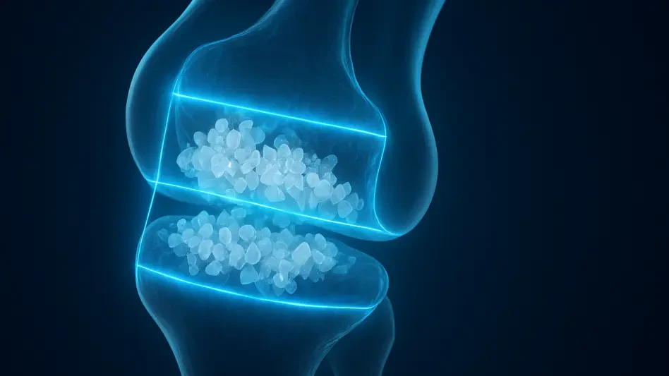

The integration of deep learning algorithms into the diagnostic workflow of rheumatology is no longer a peripheral experiment but a central necessity for achieving modern clinical precision in chronic disease management. While gout is often dismissed as a historical ailment, its contemporary prevalence is rising, and the demand for objective monitoring of tophaceous deposits has outpaced the manual capacity of human clinicians. Manual segmentation, the traditional gold standard where a sonographer meticulously outlines every millimeter of a urate crystal deposit on an ultrasound screen, is a grueling process prone to subjective bias and fatigue. The shift toward AI-based segmentation represents a fundamental pivot from qualitative “guestimation” to quantitative data science, allowing for a level of diagnostic granularity that was previously inaccessible in a high-volume clinical setting.
Automated Image Analysis in Rheumatology
The technological evolution of rheumatology has been marked by a move toward non-invasive imaging that can provide immediate, bedside feedback. Musculoskeletal ultrasound has emerged as the frontrunner in this space because it offers a radiation-free, cost-effective alternative to dual-energy CT or MRI. However, the inherent “noise” in ultrasound images—such as speckle artifacts and low contrast between different soft tissues—makes the identification of gouty tophi particularly difficult. AI-based segmentation addresses this by utilizing neural networks trained on thousands of annotated images, effectively teaching the software to recognize the specific acoustic signatures of monosodium urate crystals. This technology does not merely assist the doctor; it provides a standardized baseline that remains consistent across different clinics and practitioners.
The relevance of this technology in the current landscape cannot be overstated, as the medical community moves toward a “treat-to-target” philosophy. In this paradigm, the goal is not just to manage pain but to physically dissolve the crystal burden within the joints. To prove that a specific medication is working, clinicians need to measure the shrinking volume of a tophus over time. By automating this measurement, AI transforms ultrasound from a simple visualization tool into a sophisticated instrument for volumetric tracking. This transition is essential for validating the efficacy of urate-lowering therapies and providing patients with tangible, data-driven evidence of their recovery progress.
Core Deep Learning Architectures for Tophus Detection
Modern computer vision relies on deep neural networks that can analyze images at multiple levels of abstraction, from simple edges to complex anatomical shapes. For gout management, the challenge lies in the irregular and “fuzzy” boundaries of the tophi, which often blend into surrounding tendons or joint capsules. Unlike traditional software that uses hard-coded rules to find objects, deep learning architectures learn features directly from the data. This allows the system to adapt to the variations in image quality caused by different ultrasound machine brands or patient-specific factors like skin thickness and joint inflammation.
These architectures are generally built on convolutional layers that scan the image for specific patterns. By stacking dozens of these layers, the network can understand the context of a lesion—recognizing, for instance, that a bright spot near a bone surface is likely a tophus, whereas a similar bright spot in the middle of a muscle might be an artifact. This contextual awareness is what separates modern AI from the basic thresholding techniques of the past, providing a robust framework that can handle the inherent messiness of human anatomy and the technical limitations of real-time imaging.
Encoder-Decoder Frameworks and U-Net
The U-Net architecture remains the industry standard for medical image segmentation due to its unique symmetrical design. It functions by first “encoding” the image—compressing the spatial information into a dense vector of features to understand the “what” of the object—and then “decoding” that information back into the original image size to determine the “where.” This process ensures that the model maintains a high degree of spatial accuracy, which is critical when measuring the exact borders of a tophus. The performance of U-Net is particularly notable for its efficiency; it can process images in under 70 milliseconds, making it ideal for integration into portable, hand-held ultrasound devices.
What makes U-Net and its derivatives, such as Attention U-Net, so effective in rheumatology is their ability to prioritize relevant pixels. Attention gates within the network act as a filter, suppressing background noise and highlighting the regions of the image that contain the urate deposits. This significantly reduces the rate of false positives, which is a common hurdle in ultrasound analysis where bone shadows can often be mistaken for crystalline masses. By focusing the computational power on the most significant anatomical features, these frameworks provide a balance of speed and precision that is difficult for other models to match.
Advanced Spatial Analysis with TransUNet and Deeplabv3+
As the field matures, more complex models like TransUNet and Deeplabv3+ are being deployed to handle the most challenging diagnostic cases. TransUNet introduces the concept of self-attention mechanisms, originally developed for natural language processing, into the visual domain. This allows the model to look at the entire image at once and understand the global relationship between different structures, rather than just looking at local pixel neighborhoods. This global context is vital for segmenting large or multi-lobed tophi that might span across a wide field of view, ensuring that the AI does not lose track of the lesion’s overall structure.
In contrast, Deeplabv3+ utilizes atrous spatial pyramid pooling, a technique that allows the network to “see” at different scales simultaneously. This is particularly useful in gout management because tophi can range from microscopic grains to massive lumps several centimeters in diameter. By analyzing the image at multiple resolutions, the model can accurately capture both the fine details of small deposits and the broad boundaries of large masses. While these models require more computational power than the standard U-Net, their superior accuracy on complex datasets makes them the preferred choice for high-end diagnostic workstations and academic research where precision is more important than raw speed.
Emerging Trends in Volumetric Quantification
The most significant shift currently occurring in the field is the transition from two-dimensional area measurements to three-dimensional volumetric quantification. From 2026 to 2028, the industry expects to see a surge in software that can reconstruct a 3D model of a tophus from a series of 2D ultrasound slices. This is a game-changer for clinical trials, as volume is a much more sensitive indicator of treatment response than diameter or area. Emerging trends also indicate an move toward “automated labeling,” where the AI can automatically identify not just the size, but the density and composition of the tophus, potentially predicting how quickly it will respond to medication.
Real-World Clinical and Pharmaceutical Applications
In the clinical setting, AI-based segmentation is being used to democratize expertise. Junior clinicians can use these tools to achieve diagnostic results that are on par with senior sonographers, reducing the “learning curve” associated with musculoskeletal imaging. This ensures that a patient in a rural clinic receives the same quality of assessment as one in a major urban hospital. The technology acts as a digital safety net, flagging suspicious regions that a human observer might miss during a rushed examination, thereby increasing the overall diagnostic yield of the department.
Beyond the clinic, the pharmaceutical industry is increasingly relying on these automated tools to streamline drug development. During clinical trials for new urate-lowering drugs, thousands of ultrasound images must be analyzed to prove that the medication is effective. Using human experts for this task is prohibitively expensive and slow. AI can process these images in a fraction of the time with zero inter-observer variability, providing “cleaner” data that can lead to faster regulatory approval for life-changing treatments. This objective evidence is becoming a requirement for insurers and health systems looking for proven value in new medical interventions.
Technical Limitations and Data Heterogeneity Challenges
Despite the rapid progress, the technology faces significant hurdles regarding data heterogeneity. An AI model trained on images from a high-end stationary ultrasound machine may fail when presented with images from a budget portable device due to differences in resolution and gain settings. This “domain shift” remains a primary obstacle to widespread adoption. Furthermore, the “ground truth” used to train these models is still based on human interpretation, meaning that the AI may inadvertently learn the biases or errors of the original sonographers who annotated the initial dataset.
Ongoing development is focused on creating “vendor-neutral” algorithms that can perform consistently across any hardware platform. Researchers are also exploring semi-supervised learning techniques, where the AI can learn from unlabelled data, reducing the reliance on manual annotations. There is also the challenge of the “fuzzy boundary” problem; in some patients, the transition from tophus to healthy tissue is so gradual that even the most advanced AI struggles to find a clear edge. Addressing these edge cases requires larger, more diverse multicenter datasets that represent a broader range of patient demographics and disease stages.
The Future of AI-Driven Gout Management
Looking ahead, the next phase of development will involve the integration of AI segmentation with other diagnostic markers, such as serum uric acid levels and genomic data, to create a holistic “digital twin” of the patient. This would allow rheumatologists to run simulations on how a specific patient’s tophi might respond to different drug dosages. We are also likely to see the emergence of real-time AI guidance, where the software provides live feedback to the sonographer during the scan, ensuring that the best possible images are captured for analysis.
The long-term impact of this technology will be the total standardization of gout care. As these tools become a native feature of ultrasound hardware, the subjectivity of the “human eye” will be replaced by the precision of the “algorithmic eye.” This will shift the focus of the rheumatologist from the technical task of measurement to the strategic task of patient management. Ultimately, the automation of tophus segmentation is the first step toward a future where chronic inflammatory diseases are managed with the same level of data-driven accuracy as modern oncology or cardiology.
Conclusion: Assessing the Path to Standardized Care
The transition toward automated systems provided a necessary foundation for standardized rheumatological care, effectively removing the bottlenecks of manual labor and subjective interpretation. By successfully implementing architectures like U-Net and TransUNet, researchers established that artificial intelligence could match, and in some cases exceed, the accuracy of human specialists. The move from qualitative observation to quantitative volumetric data created a more rigorous framework for both clinical practice and drug development, ensuring that treatment efficacy was measured with mathematical certainty.
Actionable next steps should focus on the creation of a universal, multi-vendor dataset to solve the remaining issues of hardware incompatibility and data heterogeneity. The industry must move beyond isolated pilot studies and work toward integrating these AI modules directly into the firmware of portable ultrasound devices for immediate bedside utility. Furthermore, future software updates should prioritize the development of multi-modal diagnostic platforms that combine imaging data with biochemical markers. This holistic approach provided the definitive path toward not just managing gout symptoms, but systematically monitoring and achieving the complete dissolution of the crystalline burden in patients worldwide.