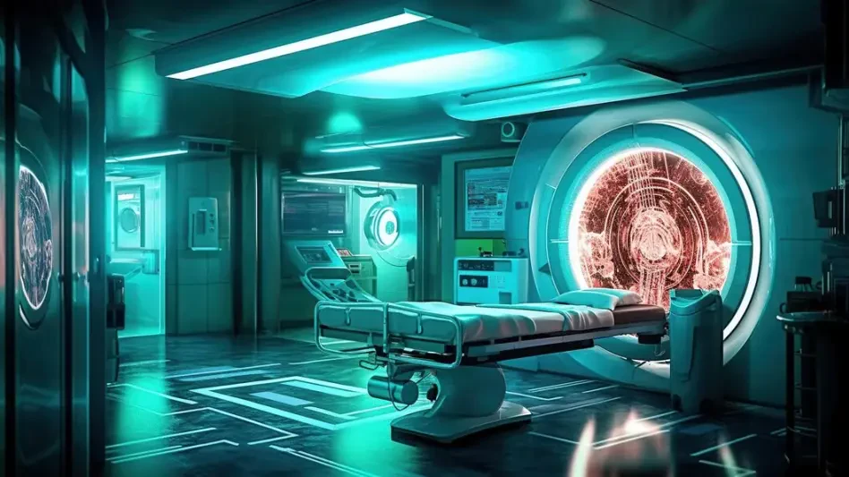

The prolonged hum and tight confinement of a traditional magnetic resonance imaging scanner have long been a source of anxiety and discomfort for patients, but a new wave of artificial intelligence is fundamentally rewriting this diagnostic experience from the inside out. The adoption of AI-powered MRI technology represents a significant advancement in diagnostic imaging and patient care. This review will explore the evolution of this technology, its key features, performance metrics, and the impact it has had on clinical applications, with a specific focus on the implementation of Siemens Healthineers’ Deep Resolve software at Penn State Health. The purpose of this review is to provide a thorough understanding of the technology, its current capabilities, and its potential for future development.
The Dawn of AI in Magnetic Resonance Imaging
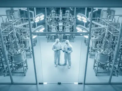
The integration of artificial intelligence into MRI was born not from technological novelty but from urgent clinical necessity. For decades, the high diagnostic value of MRI has been offset by its primary drawbacks: exceptionally long scan times that test patient endurance and increase the likelihood of motion-related image artifacts. These challenges are particularly acute in pediatric and emergency medicine, where patient cooperation is limited and speed is critical.
This persistent demand for faster, more patient-friendly imaging spurred the development of AI-driven solutions. By leveraging machine learning, developers sought to overcome the physical limitations of conventional hardware, aiming to produce high-quality diagnostic images from significantly less raw data. The goal was to create a system that could maintain or even enhance image clarity while dramatically reducing the time a patient needs to spend inside the scanner, thereby addressing the core issues of patient discomfort and operational inefficiency.
Core Technology Breakdown Deep Resolve
Deep Learning for Image Reconstruction
At the heart of the Deep Resolve software is a sophisticated deep-learning algorithm trained to reconstruct images from undersampled MRI data. Traditionally, creating a clear MRI requires collecting a vast amount of raw data, a process that is inherently time-consuming. This AI, however, has been trained on thousands of high-quality image sets, learning to identify and eliminate the noise and artifacts that typically arise when data acquisition is cut short.
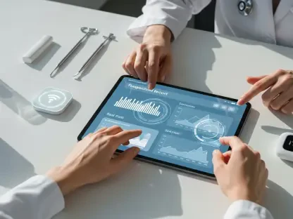
The result is a paradigm shift in image processing. Instead of a linear, data-intensive reconstruction, the AI intelligently fills in the gaps, generating images that are not only produced faster but are often sharper and clearer than those from conventional methods. This improvement in image fidelity enhances diagnostic confidence for radiologists, allowing them to make more precise assessments from scans that are far less taxing on the patient.
Accelerated Scan Acquisition Protocols
The most transformative benefit of this technology is its ability to accelerate scan acquisition. At institutions like Penn State Health, the implementation of Deep Resolve has enabled a knee MRI to be completed in as little as seven minutes, while complex brain scans have been shortened by up to 70%. This dramatic reduction in time redefines the MRI experience and optimizes hospital workflow.
These accelerated protocols have profound real-world implications. For hospitals, faster scans mean higher patient throughput, which can significantly reduce appointment backlogs and wait times. For patients, the abbreviated time in the scanner minimizes discomfort and anxiety, leading to a more positive healthcare experience and a lower probability of scans being ruined by movement, which often necessitates a repeat procedure.
Landmark Implementation at Penn State Health
Penn State Health’s system-wide adoption of Deep Resolve serves as a compelling case study for the technology’s clinical viability. Before its full rollout, the software underwent a rigorous validation process, where its AI-generated images were compared against those produced by standard, longer protocols to ensure diagnostic equivalency. The results were so conclusive that the technology was rapidly integrated into clinical practice.
Since its installation, the software is now used for approximately 90% of all pediatric MRI scans at Penn State Health Golisano Children’s Hospital, demonstrating a high level of trust from clinicians. The success in the pediatric department has also catalyzed a trend toward expanding its use to adult patient populations. This strategic rollout across eight MRI scanners in the health system is designed to streamline services, improve patient comfort, and standardize a higher level of care.
Clinical Applications and Patient Centered Impact
The most critical application of this AI-accelerated technology is in pediatric imaging, where it directly addresses the risks associated with anesthesia. Young children often require sedation to remain still for the duration of a conventional MRI scan, but their developing bodies are more vulnerable to its effects. By shortening the average exam time by seven to ten minutes, Deep Resolve significantly reduces the duration of anesthesia exposure.
This advancement has made procedures safer and less stressful for the hospital’s youngest patients and their families. In a testament to its patient-centered impact, the combination of faster scans and dedicated support from the hospital’s Child Life team has, in some cases, eliminated the need for sedation altogether. This not only enhances safety but also transforms a potentially traumatic event into a more manageable experience.
Overcoming Challenges in Clinical Adoption
Despite its clear advantages, the widespread adoption of AI-powered MRI technology is not without its challenges. The foremost hurdle is the need for meticulous validation at each institution to build clinical trust and confirm that faster scans do not compromise diagnostic accuracy. Radiologists and technologists must be confident that the AI-reconstructed images are a reliable basis for clinical decisions.
Furthermore, a system-wide rollout involves significant logistical and financial considerations. The cost of acquiring and implementing such advanced software can be substantial, though in the case of Penn State Health, this was mitigated by philanthropic contributions. Additionally, comprehensive staff training is essential to ensure that the technology’s full capabilities are leveraged effectively, transforming it from a novel tool into an integrated part of the standard of care.
The Future of AI Accelerated Diagnostics
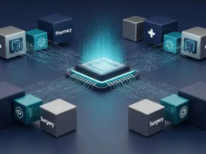
The successful implementation of technologies like Deep Resolve points toward a future where diagnostic imaging is faster, safer, and more accessible. Ongoing developments in AI are likely to yield even greater reductions in scan times, potentially making real-time imaging for dynamic processes more common. The principles of AI-driven reconstruction are also poised to expand beyond MRI to other modalities, such as CT and PET scans.
In the long term, the integration of AI is set to have a lasting impact on healthcare economics and standards. By increasing diagnostic efficiency and patient throughput, these technologies can help alleviate pressure on strained healthcare systems. As AI becomes more sophisticated, it will continue to elevate the standard of care, making advanced diagnostics a more routine and less burdensome aspect of medicine for patients globally.
Conclusion A New Standard in Medical Imaging
The adoption of AI-powered MRI technology marked a pivotal moment in the evolution of diagnostic medicine. It proved that the long-standing compromise between scan time and image quality could be effectively overcome, establishing a new clinical benchmark. The successful implementation at leading institutions confirmed the technology’s capacity to dramatically enhance patient safety, particularly by reducing anesthesia exposure in children, while simultaneously improving diagnostic clarity. Ultimately, its ability to optimize clinical operations and elevate the patient experience solidified its role as a transformative force in the field.