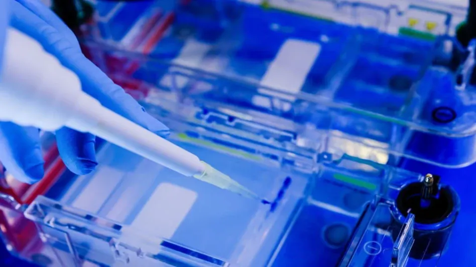
Cell sorting technologies have significantly evolved since the mid-20th century, transforming rudimentary methods into sophisticated systems fundamental to modern biological and medical research. These technologies enable the precise isolation and detailed characterization of single cells, providing invaluable insights into biological processes and disease mechanisms. This comprehensive summary will explore the historical development, current advancements, and future implications of cell sorting technologies, focusing on their applications, efficiency, and usability.
Historical Background
The Inception of Cell Sorting Technologies
The inception of cell sorting technologies dates back to 1965 when physicist Mack J. Fulwyler developed the first prototype at the Los Alamos National Laboratory. This groundbreaking invention combined a Coulter volume sensor with the recently invented ink jet printer. The initial concept was based on the principle of sorting particles by size, but Fulwyler’s approach allowed the sorting of cells based on their electrical properties, creating a new avenue for separating and analyzing biological cells. This prototype’s potential was quickly recognized by Leonard Herzenberg of Stanford University, who, with the help of engineers, modified the design to sort live cells using fluorescence. Herzenberg’s innovation revolutionized the field by introducing the ability to tag specific cell populations with fluorescent markers and sort them based on their fluorescent properties.
The early prototype provided the foundation for subsequent advancements in cell sorting technologies. Fulwyler’s and Herzenberg’s breakthroughs contributed to an entirely new way of examining cells, enabling researchers to analyze and sort cells with unprecedented precision. With these capabilities, scientists could gain deeper insights into cellular functions, facilitating research in immunology, cancer biology, and other critical fields. Overall, these initial developments marked a pivotal moment in the history of cell sorting technologies, laying the groundwork for future innovations that would continue to push the boundaries of biological and medical research.
Early Prototypes and Commercialization
Following the creation of initial prototypes, cell sorting technology saw further refinement and a push towards commercialization. Herzenberg’s team at Stanford University developed two significant early prototypes: one employing a mercury arc lamp and another using an argon ion laser to detect cells tagged with fluorescent markers. The development of these prototypes was instrumental in demonstrating the practical application of fluorescence for cell sorting and paved the way for more advanced technologies. The technology quickly gained attention, leading to the creation of the first commercial cell sorter, the Fluorescence-Activated Cell Sorter (FACS), introduced by Becton Dickinson (BD) in 1975.
The launch of the commercial FACS system marked a turning point in cell sorting technology, making it more accessible to the broader scientific community. The FACS system enabled scientists to sort and analyze cells with high precision, advancing research in various fields, from immunology to oncology. The ability to sort cells based on fluorescence opened new possibilities for studying cellular heterogeneity, analyzing immune responses, and identifying rare cell populations. As a result, FACS technology became an indispensable tool in biomedical research, laying the groundwork for future advancements in cell sorting technologies.
Key Advancements and Technologies
Microfluidic-Based Sorting
One notable advancement in cell sorting technology is the development of microfluidic-based systems. These cutting-edge platforms, such as the Bio-Techne Pala™ Cell Sorter and Pala Single-Cell Dispenser, offer several advantages over traditional cytometric-based sorters. Microfluidics provides a more streamlined and efficient approach to cell sorting, utilizing small channels to manipulate cells at the microscale. Dr. Lina Chakrabarti’s team at AstraZeneca has harnessed the Pala platform for generating monoclonal CHO cell lines used in the production of targeted recombinant protein biotherapeutics. In contrast to conventional cytometric sorters, which rely on optical and electronic methods to detect and sort cells, microfluidic-based systems utilize fluid flow techniques to separate cells, resulting in enhanced precision and speed.
The simplicity and small footprint of microfluidic systems make them particularly appealing for laboratory use. In addition to their compact size, these platforms offer increased efficiency and gentle handling of cells, which reduces stress and enhances sorting precision. The reduced cell stress is advantageous for sensitive applications where cell viability and function must be maintained. Furthermore, microfluidic systems are beneficial for applications requiring adherence to regulatory requirements, such as providing proof of monoclonality in biopharmaceutical production. Monoclonality is essential for ensuring the uniformity and consistency of therapeutic proteins, and the ability of microfluidic platforms to deliver reliable proof of monoclonality makes them invaluable tools in the pharmaceutical industry.
Benefits of Microfluidic Systems
Microfluidic systems provide numerous benefits that position them as superior alternatives to traditional cytometric-based sorters in various research applications. These systems are praised for their simplicity, as they often require less customization and fewer components than conventional sorters. This simplicity translates to lower maintenance and operational costs, making them more accessible to a wider range of laboratories. Additionally, the small footprint of microfluidic platforms allows them to fit into smaller laboratory spaces, enabling researchers to maximize their workspace efficiently. Despite their compact size, microfluidic systems can achieve high throughput and precise cell sorting, ensuring that researchers can process large cell populations accurately and swiftly.
Another significant advantage is the gentle handling of cells, which is crucial for applications where cell integrity must be preserved. Traditional cytometric sorters use high-pressure fluid systems that can introduce mechanical stress, potentially affecting cell viability and function. In contrast, microfluidic systems utilize low-pressure fluid flow techniques, minimizing mechanical stress and preserving cell integrity. This gentler approach is particularly beneficial for sorting fragile or sensitive cell types that may be adversely affected by conventional methods. As a result, microfluidic platforms are increasingly favored for applications requiring high precision and cell viability, such as single-cell genomics, stem cell research, and therapeutic cell manufacturing.
Flow Cytometry Developments
Evolution of Flow Cytometry
Flow cytometry, a core technology in cell sorting, has evolved significantly since its inception. Early flow cytometers were limited to single lasers and a few detection colors, restricting their ability to analyze complex cell populations. However, advancements in laser and detector technologies have transformed flow cytometry into a powerful and versatile tool. Modern flow cytometers, such as the Beckman Coulter® CytoFLEX SRT, offer multiple lasers and an expanded range of detectors, fluorochromes, and antibodies, enabling the simultaneous analysis and sorting of multiple cell populations. These improvements have significantly enhanced the capabilities of flow cytometry, allowing researchers to investigate cellular heterogeneity and complex biological processes with greater accuracy.
In addition to hardware advancements, software capabilities have also seen substantial improvements. Sophisticated software interfaces enable researchers to design complex experiments, analyze data more efficiently, and automate various processes, increasing workflow productivity. These advancements have made flow cytometry more accessible to a broader range of users, making it possible for even novice researchers to operate the instruments after brief training. The integration of biohazard protection features has also made flow cytometry safer, allowing researchers to work with potentially hazardous samples with confidence. Overall, these developments have cemented flow cytometry’s position as a cornerstone technology in cell analysis and sorting.
Modern Flow Cytometry Instruments
Modern flow cytometry instruments, such as the CytoFLEX SRT, exemplify the advancements in this technology. The CytoFLEX SRT is favored for its simplicity and flexibility, enabling researchers to perform complex experiments with minimal training. The instrument’s user-friendly interface and automated processes streamline the workflow, allowing researchers to focus on their experiments rather than instrument operation. This level of automation is critical for extensive and continuous research operations, where efficiency and reliability are paramount. Automated processes, such as sample loading, data acquisition, and data analysis, reduce the likelihood of human error and improve the overall accuracy of experiments.
Another notable feature of modern flow cytometry instruments is their ability to handle multiple populations simultaneously. The expanded range of detectors and fluorochromes allows researchers to label and analyze various cell populations within a single experiment, providing a comprehensive view of cellular heterogeneity. This capability is particularly valuable in fields such as immunology, oncology, and stem cell research, where understanding the diversity and complexity of cell populations is crucial. Despite the advent of newer instruments, older models like the BD FACSAria™ remain in use due to their established reliability and user familiarity. The continued use of these legacy instruments demonstrates the enduring value of flow cytometry in scientific research, underscoring the importance of ongoing innovation and development in this field.
Spectral Analysis and Real-Time Imaging
Integration of Spectral Analysis
A significant leap in cell sorting technology is the integration of spectral analysis and real-time imaging, exemplified by the BD FACSDiscover™ S8. Spectral analysis allows researchers to differentiate and sort cells based on their spectral properties, providing a more nuanced understanding of cell populations. Real-time imaging enhances this capability by enabling researchers to visualize cells as they are being sorted, offering immediate feedback and facilitating more precise sorting decisions. This technology combines deep multiparametric phenotyping with real-time imaging, allowing researchers to categorize and sort rare cell populations based on internal marker localization. The ability to simultaneously analyze multiple parameters at once can streamline workflows and improve accuracy, particularly in complex applications such as tumor analysis.
The integration of spectral analysis and real-time imaging is particularly advantageous for identifying and sorting rare cell populations. Traditional sorting methods may struggle with low-abundance cells or those that exhibit subtle differences in marker expression. Spectral analysis enhances the resolution and sensitivity of cell sorting, making it possible to detect and isolate these elusive populations. Real-time imaging complements spectral analysis by providing visual confirmation of cell characteristics, reducing the risk of sorting errors and increasing confidence in the results. Together, these technologies offer a powerful approach to cell sorting, enabling researchers to tackle challenging applications with greater precision and efficiency.
Applications in Cancer Research
The spectral performance of technologies like the BD FACSDiscover S8 facilitates complex multiparametric applications, enhancing the ability to analyze and sort cells with high precision. It has proven particularly valuable in cancer research, where understanding tumor progression and cellular heterogeneity is crucial. Tumors are composed of diverse cell populations, each with distinct genetic and phenotypic characteristics. Traditional sorting methods may overlook these subtle differences, but the advanced capabilities of modern cell sorters allow for the detailed analysis of complex samples, enabling researchers to isolate specific subpopulations for further study. By identifying and analyzing these unique cell populations, researchers can gain insights into tumor biology, identify potential therapeutic targets, and develop more effective treatments.
Consistency and reliability of instruments like the BD FACSDiscover S8 ensure that observed differences in cell populations stem from biological variability rather than instrumental drift. This consistency is crucial for reproducibility, a fundamental aspect of scientific research. Researchers can trust that their results are accurate and representative, allowing them to build on their findings with confidence. The ability to sort and analyze rare cell populations with high precision also opens new avenues for cancer research, such as studying circulating tumor cells, investigating tumor microenvironments, and exploring the mechanisms of resistance to cancer therapies. Overall, the integration of spectral analysis and real-time imaging in cell sorting technologies represents a significant advancement, offering powerful tools for advancing cancer research and improving patient outcomes.
Challenges and Future Directions
Current Challenges
Despite the remarkable advances, cell sorting technologies still face challenges that must be addressed to fully realize their potential. One of the primary challenges is the high cost and complexity of instruments. State-of-the-art cell sorters can be prohibitively expensive for some laboratories, particularly smaller or resource-limited facilities. The initial investment and ongoing maintenance costs may limit access to these advanced technologies, potentially hindering scientific progress. Additionally, the complexity of operating and maintaining these instruments requires specialized training and expertise, which may not be readily available in all research settings.
Another challenge is the need for continuous development to keep pace with the increasing demand for more sophisticated analysis. As researchers delve deeper into the complexities of biological systems, the requirements for precision, throughput, and scalability continue to rise. Current technologies, while highly advanced, may still fall short in meeting these evolving demands. For example, the sorting and analysis of extremely rare cell populations, such as circulating tumor cells, require even higher sensitivity and accuracy. Addressing these challenges will necessitate ongoing innovation and collaboration between researchers, engineers, and technology developers.
Future Improvements
Looking ahead, several potential improvements could enhance the accuracy and efficiency of cell sorting technologies. One promising avenue is the integration of clustering algorithms and artificial intelligence (AI) for automated identification and tagging of specific cell populations. AI-powered algorithms can analyze large datasets, identify patterns, and make predictions in real-time, streamlining the sorting process and reducing the reliance on manual intervention. By leveraging machine learning techniques, these systems can continuously improve their accuracy and adapt to new challenges, providing increasingly precise sorting capabilities.
Other future advancements could involve the development of more portable and cost-effective instruments. Reducing the size and cost of cell sorters would make them more accessible to a broader range of laboratories, enabling more researchers to benefit from advanced cell sorting technologies. Miniaturized and affordable systems could also facilitate field-based research, expanding the applications of cell sorting to new environments and populations. Additionally, the integration of advanced imaging techniques and multiplexed detection methods could further enhance the capabilities of cell sorting technologies, enabling researchers to analyze and sort cells with even greater resolution and specificity.
Conclusion
Since the mid-20th century, cell sorting technologies have undergone significant evolution, transitioning from basic techniques to intricate systems crucial for contemporary biological and medical research. These advanced methodologies allow for the precise isolation and detailed analysis of individual cells, offering deep insights into biological processes and disease mechanisms. This comprehensive overview will delve into the historical development, current innovations, and future prospects of cell sorting technologies, concentrating on their range of applications, efficiency, and ease of use.
Initially, cell sorting was a rudimentary process, but technological advancements have transformed it into a highly sophisticated practice. Modern cell sorting methods, such as flow cytometry and magnetic-activated cell sorting, enable researchers to identify and isolate specific cell populations with high precision. These techniques are essential for numerous applications, including cancer research, immunology, and stem cell research.
The efficiency of cell sorting technologies has substantially improved over the years, with current systems offering rapid and accurate sorting of millions of cells in a short time. This has bolstered their usability in both research and clinical settings. As technology continues to advance, the future of cell sorting holds promise for even more refined methods, potentially leading to groundbreaking discoveries in understanding diseases and developing new therapeutic approaches.


