
Evidence suggests that the timing of imaging data collection is critical, as features captured after treatment carry more weight in predicting pathological complete response. In the rapidly evolving landscape of oncology, clinicians often struggle to determine the effectiveness of intensive neoadjuvant therapies for head and neck squamous cell carcinoma before definitive surgery. Traditional imaging methods frequently fail to capture the subtle biological shifts occurring within the tumor microenvironment during treatment. This diagnostic gap can lead to unnecessary procedures or delayed adjustments in care. However, a recent multicenter study involving over five hundred patients has introduced a longitudinal machine learning framework known as the Delta-Habitat model. This computational tool specifically focuses on the spatial distribution of cancer cells and their surrounding tissue. By moving beyond static images, this approach offers a dynamic window into how malignant structures transform.
Technical Innovation: Integrating Spatial Habitat Radiomics
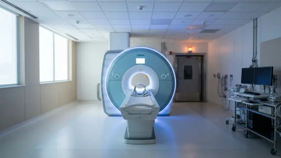
The Delta-Habitat model represents a significant technical departure from conventional delta-radiomics by analyzing distinct biological regions within a tumor rather than viewing the mass as a uniform entity. By segmenting magnetic resonance imaging data into various biological habitats, the framework can track how specific sub-regions respond to chemoimmunotherapy. This granular analysis allows the algorithm to identify patterns of resistance or sensitivity that are invisible to the naked eye. In a retrospective analysis conducted across multiple medical centers from 2026 to 2027, researchers validated that these spatial features provide a much more nuanced understanding of tumor evolution. The model demonstrated remarkable consistency, achieving an area under the curve of approximately 0.82 across diverse patient cohorts. Unlike previous iterations of predictive software, this system maintains its accuracy even when faced with the inherent variability of imaging equipment.
One of the most striking findings from this technological advancement is the discovery that post-treatment imaging data serves as the primary driver for successful predictions. While pre-treatment baseline scans provide a starting point, the structural changes observed after the initial rounds of therapy are far more indicative of long-term success. Furthermore, the study integrated clinical biomarkers such as the post-treatment neutrophil-to-lymphocyte ratio to see if biological markers could further sharpen the model’s foresight. Interestingly, while the ratio proved to be a strong independent predictor of a pathological complete response, the standalone Delta-Habitat framework remained powerful enough to function without heavy reliance on traditional blood work. This autonomy suggests that the visual data encoded in magnetic resonance imaging contains a wealth of untapped information regarding the immune system’s interaction with the cancer throughout the therapy cycle.
Clinical Integration: Enhancing Human Expertise Through Algorithms
Beyond its capacity for raw data processing, the Delta-Habitat model has proven to be a transformative tool for medical professionals in clinical environments. When compared directly against experienced radiologists, the algorithm consistently delivered more accurate predictions of how patients would respond to their specific treatment regimens. However, the true value of the technology lies in its role as a collaborative decision-support system. During testing, the inclusion of the AI’s insights significantly elevated the diagnostic performance of practicing clinicians across all experience levels. Senior radiologists experienced a notable boost in their predictive accuracy, but the most dramatic improvements were observed among junior residents. These less experienced doctors saw their diagnostic capabilities increase by nearly twenty percent when using the model’s outputs as a guide. This democratization of expertise ensures that high-quality, data-driven assessments are available.
The implementation of this longitudinal machine learning framework established a new benchmark for personalized oncology by providing a verifiable metric for treatment efficacy. It was observed that the model’s ability to categorize tumor heterogeneity allowed for more targeted therapeutic adjustments, potentially sparing patients from the toxic side effects of ineffective drugs. Moving forward, healthcare systems should prioritize the integration of spatial habitat analysis into standard diagnostic pipelines to maximize the benefits of chemoimmunotherapy. Standardizing imaging protocols to capture these post-treatment habitats will be essential for refining patient selection in future clinical trials. By leveraging these computational insights, medical teams successfully transitioned from reactive care to a more proactive, individualized strategy. The study concluded that the fusion of machine learning with traditional radiology provided a sustainable roadmap for digital oncology.